. 2010 Jun 3;6(6):e1000934.
doi: 10.1371/journal.ppat.1000934.

Epithelial p38alpha controls immune cell recruitment in the colonic mucosa

Affiliations

Epithelial p38alpha controls immune cell recruitment in the colonic mucosa

Young Jun Kang et al. PLoS Pathog. .

Abstract

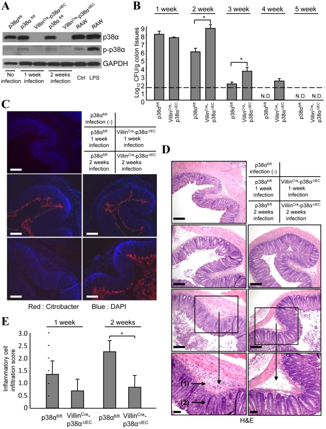
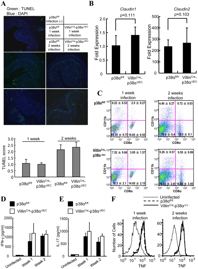
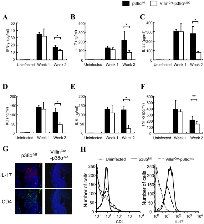
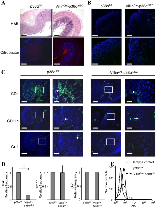
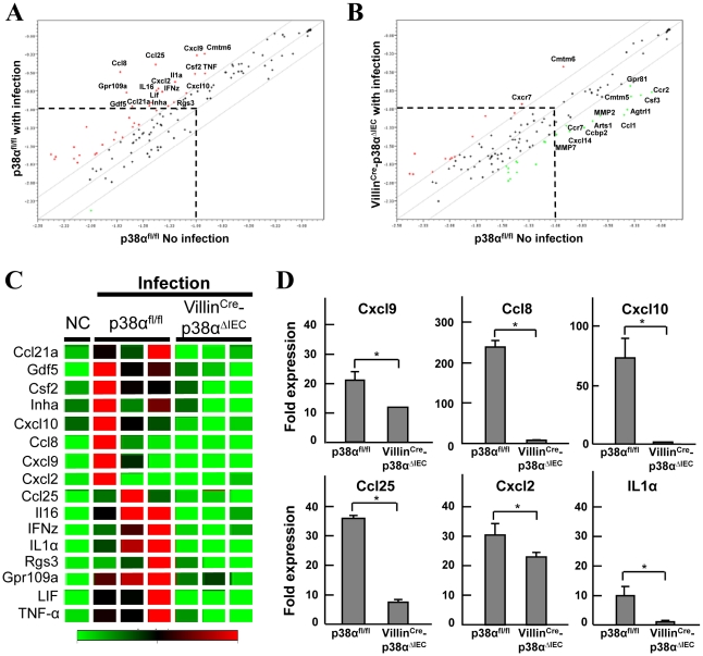
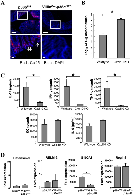
Intestinal epithelial cells (IECs) compose the first barrier against microorganisms in the gastrointestinal tract. Although the NF-kappaB pathway in IECs was recently shown to be essential for epithelial integrity and intestinal immune homeostasis, the roles of other inflammatory signaling pathways in immune responses in IECs are still largely unknown. Here we show that p38alpha in IECs is critical for chemokine expression, subsequent immune cell recruitment into the intestinal mucosa, and clearance of the infected pathogen. Mice with p38alpha deletion in IECs suffer from a sustained bacterial burden after inoculation with Citrobacter rodentium. These animals are normal in epithelial integrity and immune cell function, but fail to recruit CD4(+) T cells into colonic mucosal lesions. The expression of chemokines in IECs is impaired, which appears to be responsible for the impaired T cell recruitment. Thus, p38alpha in IECs contributes to the host immune responses against enteric bacteria by the recruitment of immune cells.


Conflict of interest statement

The authors have declared that no competing interests exist.

Figures

Figure 1
Figure 1. p38α in intestinal epithelial cells is required for the immunity to C. rodentium.
A, The level of p38α and phosphorylated-p38α (p-p38α) in isolated IECs from non-infected mice (lane 1), or p38αfl/fl and VillinCre-p38αΔIEC mice infected with C. rodentium for 1 or 2 weeks (lane 2, 4 and 3, 5). RAW264.7 cells, a mouse macrophage cell line, treated with or without LPS for 1 hour were used as controls for p38α phosphorylation (lane 6, 7). The data is representative from two independent experiment sets. B, C. rodentium CFU recovered from colon tissues of individual p38αfl/fl and VillinCre-p38αΔIEC mice at 1, 2, 3, 4 and 5 weeks after inoculation. The data shown are from one experiment, representative of five. The transverse bar is the detection limit. Asterisk, p<0.05. Error bars indicate s.d. (n = 6). N.D. not detected. C, Immunofluorescence staining of C. rodentium in the colon segments by anti-C. rodentium antibodies (red). Nuclei were counterstained with DAPI (blue). Colon segments isolated from a non-infected p38αfl/fl control mouse, and from p38αfl/fl or VillinCre-p38αΔIEC mice at 1 and 2 weeks after infection were used. Scale bar, 100 µm. D, Inflammation detected by hematoxylin/eosin staining of colon segments. The sections are adjacent to those in C. Scale bar, 100 µm. E, The inflammatory cell infiltration into the submucosa in 1 or 2 weeks infected p38αfl/fl or VillinCre-p38αΔIEC mice was evaluated by histological score.
Figure 2
Figure 2. Epithelial integrity and immune cells are normal in VillinCre-p38αΔIEC mice after C. rodentium infection.
A, TUNEL staining (green) on colon cross sections from non-infected p38αfl/fl control and p38αfl/fl or VillinCre-p38αΔIEC mice at 1 and 2 weeks after infection. Scale bar, 100 µm. The sections were adjacent to those in Fig. 1C. B, Expression of tight junction proteins in the intestinal epithelial cells of C. rodentium-infected mice. Expression of Claudin-1 and -2 was measured by qPCR method. Fold expression of proteins was calculated over the expression of proteins of uninfected IECs. GADPH levels was measured as an internal control. C, Composition of the dendritic cell compartment in the mesenteric lymph node cells of p38αfl/fl or VillinCre-p38αΔIEC mice at 1 and 2 weeks after infection. D & E, C. rodentium-specific IFN-γ (D) and IL-17 (E) responses after restimulation of mesenteric lymph node cells from p38αfl/fl or VillinCre-p38αΔIEC mice at 1 and 2 weeks after infection. Data are representative of three independent experiments (n = 3). Cells from uninfected mice were used as a control. F, Expression of TNF-α by the macrophages (F4/80 positive cells) of the draining mesenteric lymph nodes of p38αfl/fl or VillinCre-p38αΔIEC mice at 1 and 2 weeks after infection. Data are representative of two independent experiments (n = 3).
Figure 3
Figure 3. Cytokine expressions in the colon of VillinCre-p38αΔIEC mice are impaired after C. rodentium infection.
A–F, ex vivo colon culture ELISA of IFN-γ, IL-17, IL-22, KC, IL-6, and TNF expression in p38αfl/fl or VillinCre-p38αΔIEC mice colons at 1 and 2 weeks after C. rodentium infection. Asterisk, p<0.05; Double asterisk, p>0.05. Error bars indicate s.d. Data are representative of two independent experiments (n = 3). G & H, Expression of IL-17 in the colon mucosa of p38αfl/fl or VillinCre-p38αΔIEC mice at 2 weeks after C. rodentium infection, determined by immunofluorescent staining for CD4 (green) and IL-17 (red), and nuclei were counterstained with DAPI (blue) (G), and by FACS analysis of whole colon cells using anti-CD4-PE and IL-17-APC antibodies (H). The basal levels of CD4 and IL-17 of uninfected p38αfl/fl colon cells are shown in the FACS analysis.
Figure 4
Figure 4. p38α in intestinal epithelial cells is required for chemokine expression and to recruit immune cells into the colon mucosa after C. rodentium infection.
A, Histological changes with inflammation (upper) by hematoxylin/eosin staining and the immunofluorescence staining of C. rodentium (lower; red) in the adjacent distal colon cross sections from p38αfl/fl or VillinCre-p38αΔIEC mice at 2 weeks after infection. Scale bar, 100 µm. B, CD4+ T cell infiltration in the distal colon mucosa of non-infected p38αfl/fl or VillinCre-p38αΔIEC mice, determined by immunofluorescent staining (green). Scale bar, 100 µm (low magnification; upper panels), 30 µm (high magnification; lower panels). Nuclei were counterstained with DAPI (blue). C, Cell infiltration in the distal colon mucosa of p38αfl/fl or VillinCre-p38αΔIEC mice at 2 weeks after infection, determined by immunofluorescent staining for CD4, CD11c, and Gr-1 (green). Right panels are enlarged images of the parts denoted in the boxes in the left panels in each group. Scale bar, 100 µm (low magnification), 30 µm (high magnification). D, Relative frequencies of CD4+, CD11c+, and Gr-1+ cells in the distal colon mucosa from infected VillinCre-p38αΔIEC mice in comparison with that from infected p38αfl/fl mice. Immunostained cells were counted in total ten fields of each mouse. The relative frequencies were calculated after adjusting the number from infected p38αfl/fl mice as 1. The results show mean ± s.d. from four animal pairs. Data are representative of 2–4 independent experiments. Asterisk, p<0.05. E, Infiltration of CD4+ T cells to the lamina propria. Lamina propria lymphocytes were obtained from p38αfl/fl or VillinCre-p38αΔIEC mice at 2 weeks after infection, and stained with anti-CD3-FITC and anti-CD4-PE antibodies for FACS analysis. The number of CD4+ cells is shown in the CD3+ T cell population. 5000 cells were analyzed per sample.
Figure 5
Figure 5. p38α in intestinal epithelial cells is required for chemokine expression to recruit immune cells into the colon mucosa after C. rodentium infection.
A, B, Linear scatter plot of gene expression. Each gene in the microarray is represented by a point with coordinates consisting of average gene expression in log scale (n = 3) from isolated IECs of p38αfl/fl (A) or VillinCre-p38αΔIEC (B) mice at 1 week after infection, in comparison with those of a non-infected p38αfl/fl mouse. The genes with more than a 2 fold increase or less than a 0.5 fold decrease are represented as red or green, respectively. Gene names are indicated only if those expression levels are higher than a threshold and significantly changed. C, The genes determined as induced more than 2 fold in the IECs of infected p38αfl/fl mice in (a) are selectively shown in the clustering image. D, Quantitative RT-PCR of gene expression in isolated IECs of p38αfl/fl or VillinCre-p38αΔIEC mice at 1 week after infection. Values represent amounts relative to that of uninfected p38αfl/fl mouse samples. Asterisk, p<0.05. Error bars indicate s.d. Data is representative of three experiments.
Figure 6
Figure 6. Analysis of chemokines Ccl25 and Cxcl10, and antimicrobial gene expression.
A, Ccl25 expression in the colon mucosa of uninfected p38αfl/fl and p38αfl/fl or VillinCre-p38αΔIEC mice at 1 week after infection, determined by immunofluorescent staining (red). Nuclei were counterstained with DAPI (blue). Data are representative of three independent experiments. B, Impaired bacteria clearance in C. rodentium-infected Cxcl10 knockout mice. Colon tissues from 2-week C. rodentium-infected wildtype or Cxcl10 knockout mice were obtained and bacterial CFU was determined. Asterisk, p<0.05. Error bars indicate s.d. (n = 3). C, ex vivo colon culture ELISA of IL-17, IFN-γ, TNF-α, KC, and IL-6 of C. rodentium-infected wildtype or Cxcl10 knockout mice. Asterisk, p<0.01. Error bars indicate s.d. (n = 3). D, Quantitative real-time PCR of antimicrobial gene expression in isolated IECs of p38αfl/fl or VillinCre-p38αΔIEC mice at 1 week after infection. Asterisk, p<0.05. Error bars indicate s.d. Data is representative of three experiments.

Similar articles

Cited by

References

    1. Kaper JB, Nataro JP, Mobley HL. Pathogenic Escherichia coli. Nat Rev Microbiol. 2004;2:123–140. - PubMed
    1. Mead PS, Griffin PM. Escherichia coli O157:H7. Lancet. 1998;352:1207–1212. - PubMed
    1. Borenshtein D, McBee ME, Schauer DB. Utility of the Citrobacter rodentium infection model in laboratory mice. Curr Opin Gastroenterol. 2008;24:32–37. - PubMed
    1. Eckmann L. Animal models of inflammatory bowel disease: lessons from enteric infections. Ann N Y Acad Sci. 2006;1072:28–38. - PubMed
    1. Bry L, Brigl M, Brenner MB. CD4+-T-cell effector functions and costimulatory requirements essential for surviving mucosal infection with Citrobacter rodentium. Infect Immun. 2006;74:673–681. - PMC - PubMed

Publication types

MeSH terms