. 2010 Jun 3;6(6):e1000925.
doi: 10.1371/journal.ppat.1000925.

The SOCS-box of HIV-1 Vif interacts with ElonginBC by induced-folding to recruit its Cul5-containing ubiquitin ligase complex

Affiliations

The SOCS-box of HIV-1 Vif interacts with ElonginBC by induced-folding to recruit its Cul5-containing ubiquitin ligase complex

Julien R C Bergeron et al. PLoS Pathog. .

Abstract

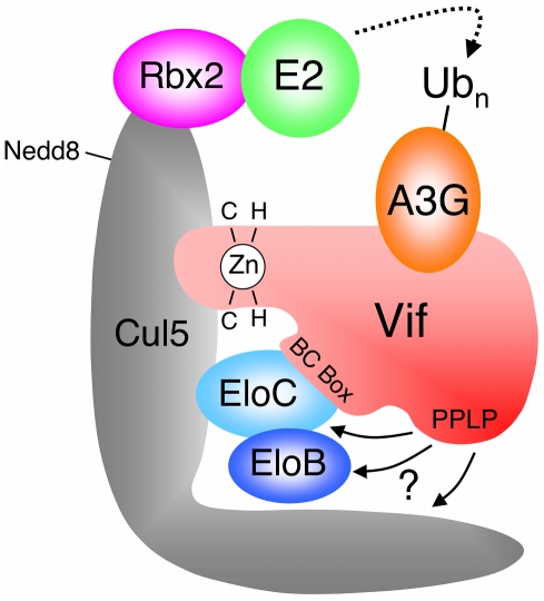
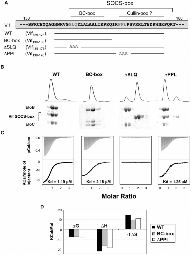
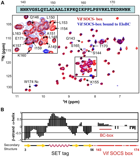
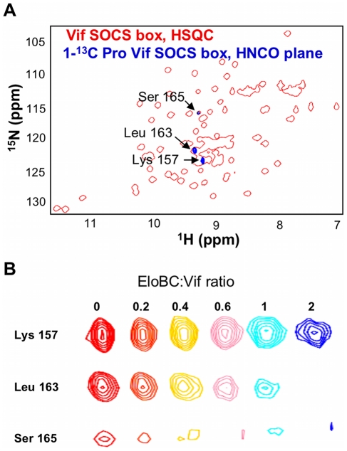
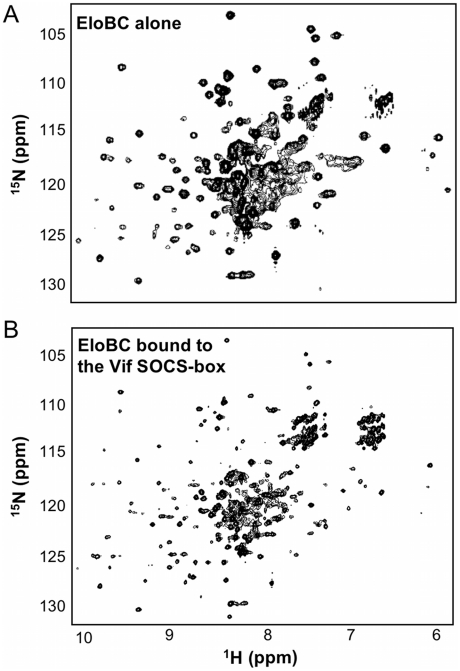
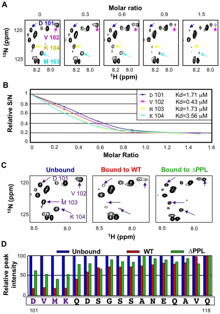
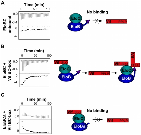
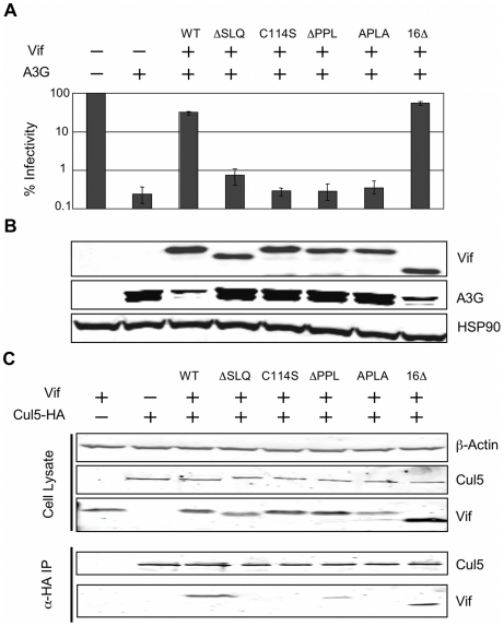
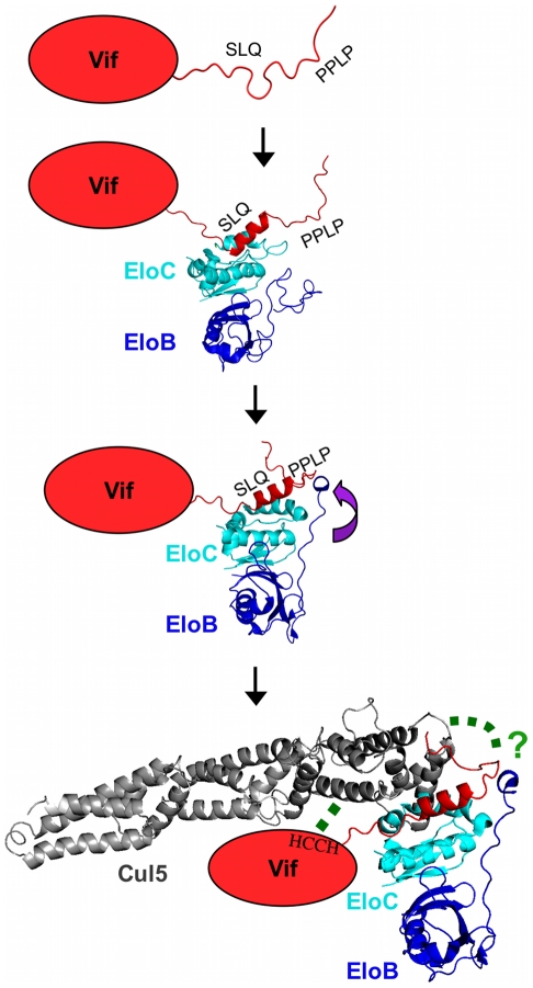
The HIV-1 viral infectivity factor (Vif) protein recruits an E3 ubiquitin ligase complex, comprising the cellular proteins elongin B and C (EloBC), cullin 5 (Cul5) and RING-box 2 (Rbx2), to the anti-viral proteins APOBEC3G (A3G) and APOBEC3F (A3F) and induces their polyubiquitination and proteasomal degradation. In this study, we used purified proteins and direct in vitro binding assays, isothermal titration calorimetry and NMR spectroscopy to describe the molecular mechanism for assembly of the Vif-EloBC ternary complex. We demonstrate that Vif binds to EloBC in two locations, and that both interactions induce structural changes in the SOCS box of Vif as well as EloBC. In particular, in addition to the previously established binding of Vif's BC box to EloC, we report a novel interaction between the conserved Pro-Pro-Leu-Pro motif of Vif and the C-terminal domain of EloB. Using cell-based assays, we further show that this interaction is necessary for the formation of a functional ligase complex, thus establishing a role of this motif. We conclude that HIV-1 Vif engages EloBC via an induced-folding mechanism that does not require additional co-factors, and speculate that these features distinguish Vif from other EloBC specificity factors such as cellular SOCS proteins, and may enhance the prospects of obtaining therapeutic inhibitors of Vif function.


Conflict of interest statement

The authors have declared that no competing interests exist.

Figures

Figure 1
Figure 1. HIV-1 Vif forms an E3 complex to promote the degradation of A3G.
Schematic representation of the E3 ubiquitin ligase complex formed by HIV-1 Vif, with motifs reported to be required for the interaction with Cul5 and EloBC indicated. Adapted from reference .
Figure 2
Figure 2. Biochemical analysis of the Vif-EloBC interaction.
(A) Schematic representation of the Vif SOCS-box constructs and mutants used in this study. The amino acid sequence is indicated on top, with the mutated residues in gray. (B) Gel filtration binding assay. The Vif SOCS-box and mutated variants were mixed with equimolar amounts of EloBC and run through a gel filtration column, with the UV280 trace shown on top. The eluted fractions were collected and run on a 16% polyacrylamide gel and stained with Coomassie blue (bottom). (C) ITC binding assay. The Vif fusion proteins were titrated against EloBC. The raw data are shown on top, the heat integration at the bottom. The resulting Kd is given for each construct, except for the ΔSLQ protein, for which no binding was observed. (D) Thermodynamic analysis of the ITC binding assay. The binding free energy (ΔG), enthalpy (ΔH) and entropy (ΔS) are plotted for the Vif fusions proteins binding to EloBC. The ΔSLQ protein is not shown as no binding was observed.
Figure 3
Figure 3. NMR spectroscopy of the Vif SOCS-box.
(A) 15N-HSQC spectrum of Vif SOCS-box protein in the free state (red spectrum) and bound to EloBC (blue spectrum), with the sequence of the Vif SOCS-box shown on top. The peaks assigned to residues belonging to the Vif SOCS-box are indicated; some peaks are omitted for clarity. (B) Secondary structure of the Vif SOCS-box wild type protein, measured with the software SSP using chemical shifts values. The portion of this protein corresponding to the Vif SOCS-box is indicated in red, with the location of the BC-box indicated. The numbering indicated below corresponds to the SET tag (black) and the Vif sequence (red).
Figure 4
Figure 4. Proline labelling of the Vif SOCS-box protein.
(A) NH plane of the HNCO spectrum collected on 15N, 1-13C proline-labelled Vif SOCS-box protein (blue spectrum), overlapped with the 15N-HSQC spectrum of this protein (red spectrum). (B) Titration of EloBC against proline-labelled Vif SOCS-box, with the ratio of EloBC to Vif shown on top. The peak for Leu 163 disappears upon binding, but the peak for Lys 157 is not affected. The peak for Ser 165 also decreases in intensity, and is shifted upfield on both proton and nitrogen dimensions.
Figure 5
Figure 5. NMR spectroscopy of EloBC.
15N-HSQC spectrum of protonated EloBC (A) or uniformly-deuterated EloBC bound to unlabelled wild type Vif SOCS-box protein (B). The best spectra available were used for illustration, but similar spectra were recorded on several occasions using different protein preparations. The spectrum of free EloBC shows some well dispersed peaks, but also significant overlap exists between 7.5 and 8.5 ppm in the 1H dimension, indicative of an unstructured domain. The spectrum of bound EloBC is much better resolved, with more peaks corresponding to well-folded domains.
Figure 6
Figure 6. The PPLP motif of Vif interacts with residues 101–104 of EloB.
(A) Unlabelled Vif SOCS box protein was titrated against 15N-labelled EloBC, and 15N-HSQC spectra were recorded for each point of the titration. For these spectra, the contrast is set in order to visualize only the very intense peaks assigned to the C-terminus of EloB. (B) The relative intensities of the peaks assigned to residues 101–104 of EloB were fitted to corresponding Kd values using a script written with the Mathematica software. The Kd values thus obtained are comparable to the values obtained by ITC. (C) 15N-HSQC NMR spectrum of EloBC alone (left panel), bound to the wild type Vif SOCS-box protein (middle panel) or to the ΔPPL protein (right panel); these spectra are shown with a low contrast in order to visualize only the very intense peaks. (D) For each peak assigned to the C-terminus of EloB, the S/N was measured using SPARKY and plotted as a relative intensity compared to the S/N of peaks in the unbound protein.
Figure 7
Figure 7. ITC measurement of the Vif-EloB interaction.
For all panels, ITC data is shown on the left, with the raw data on top and the heat integration at the bottom. A schematic representation of the experiment is shown on the right. (A) Titration of EloBC complex against the ΔSLQ protein; no binding is observed. (B) Titration of the EloBC/Vif BC-box complex against the ΔSLQ protein. (C) Titration of the EloBCΔ/Vif BC-box complex against the ΔSLQ protein; no binding is measured, but an endothermic reaction is observed instead.
Figure 8
Figure 8. The interaction between the PPLP motif of Vif and EloB is necessary for binding to Cul5.
(A) The activity of Vif proteins was measured by the capacity to overcome the anti-viral action of A3G in a single-cycle infectivity assay. The data are represented as relative β-galactosidase units normalised to HIV-1 with no A3G or Vif. The means of three experiments was used, with error bars representing the standard deviation. (B) Immunoblot of the whole cell lysates from the producer cells used for infectivity assays. HSP90 is shown as a loading control. (C) Co-immunioprecipitation of Vif with Cul5. 293T cells were transfected with plasmids expressing HA-tagged Cul5 and wild type or mutated Vif proteins, and Cul5-HA was isolated using an α-HA antibody. Cell lysates and immunoprecipitates were analysed by immunoblotting for presence of Vif and Cul5-HA.
Figure 9
Figure 9. Schematic representation of the induced-folding mechanism for the formation of Vif-EloBC-Cul5 complex.
Structure-based representation of the Vif-EloBC-Cul5 complex formation; for simplicity, the various binding events are shown in succession, although in the absence of kinetic data the precise order of events remains to be verified. Vif is represented by a red oval apart from the SOCS-box, which was modelled on the crystal structure of the EloBC-Vif BC-box complex (PDB ID:3DCG). The structure of EloBC bound to the BC-box was modelled on the NMR structure of EloBC bound to the SOCS3 BC-box (PDB ID: 2JZ3). The structure of EloBC bound to the Vif BC-box and the PPLP motif was modelled on the crystal structure of the EloBC-SOCS2 complex (PDB ID:2C9W). The structure of Cul5 was modelled on the crystal structure of Cul1 (PDB ID:1LDJ). The green dashed lines indicate interactions between HIV-1 Vif and Cul5.

Similar articles

Cited by

References

    1. Petroski MD, Deshaies RJ. Function and regulation of cullin-RING ubiquitin ligases. Nat Rev Mol Cell Biol. 2005;6:9–20. - PubMed
    1. Willems AR, Schwab M, Tyers M. A hitchhiker's guide to the cullin ubiquitin ligases: SCF and its kin. Biochim Biophys Acta. 2004;1695:133–170. - PubMed
    1. Mahrour N, Redwine WB, Florens L, Swanson SK, Martin-Brown S, et al. Characterization of Cullin-box sequences that direct recruitment of Cul2-Rbx1 and Cul5-Rbx2 modules to Elongin BC-based ubiquitin ligases. J Biol Chem. 2008;283:8005–8013. - PubMed
    1. Kamura T, Maenaka K, Kotoshiba S, Matsumoto M, Kohda D, et al. VHL-box and SOCS-box domains determine binding specificity for Cul2-Rbx1 and Cul5-Rbx2 modules of ubiquitin ligases. Genes Dev. 2004;18:3055–3065. - PMC - PubMed
    1. Bullock AN, Debreczeni JE, Edwards AM, Sundstrom M, Knapp S. Crystal structure of the SOCS2-elongin C-elongin B complex defines a prototypical SOCS box ubiquitin ligase. Proc Natl Acad Sci U S A. 2006;103:7637–7642. - PMC - PubMed

Publication types

MeSH terms