. 2010 Jun 3;6(6):e1000927.
doi: 10.1371/journal.ppat.1000927.

A Staphylococcus aureus small RNA is required for bacterial virulence and regulates the expression of an immune-evasion molecule

Affiliations

A Staphylococcus aureus small RNA is required for bacterial virulence and regulates the expression of an immune-evasion molecule

Svetlana Chabelskaya et al. PLoS Pathog. .

Abstract

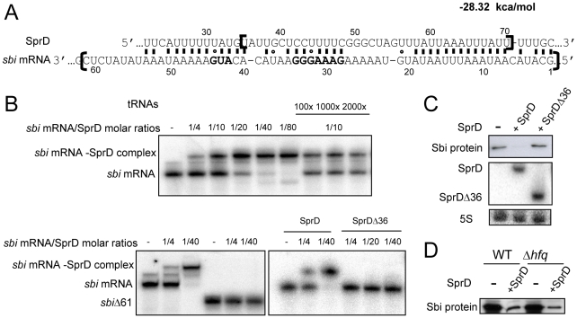
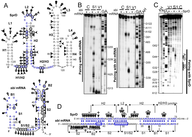
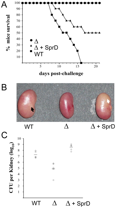
Staphylococcus aureus, a pathogen responsible for hospital and community-acquired infections, expresses many virulence factors under the control of numerous regulatory systems. Here we show that one of the small pathogenicity island RNAs, named SprD, contributes significantly to causing disease in an animal model of infection. We have identified one of the targets of SprD and our in vivo data demonstrate that SprD negatively regulates the expression of the Sbi immune-evasion molecule, impairing both the adaptive and innate host immune responses. SprD interacts with the 5' part of the sbi mRNA and structural mapping of SprD, its mRNA target, and the 'SprD-mRNA' duplex, in combination with mutational analysis, reveals the molecular details of the regulation. It demonstrates that the accessible SprD central region interacts with the sbi mRNA translational start site. We show by toeprint experiments that SprD prevents translation initiation of sbi mRNA by an antisense mechanism. SprD is a small regulatory RNA required for S. aureus pathogenicity with an identified function, although the mechanism of virulence control by the RNA is yet to be elucidated.


Conflict of interest statement

The authors have declared that no competing interests exist.

Figures

Figure 1
Figure 1. SprD RNA expression profiles in several S. aureus strains.
The expression of SprD during a 24-hour growth of S. aureus N315 (A), MRSA252 (B), RN1 (C) and RN1 ΔRNAIII (D) strains by Northern blots using labeled DNA probes for SprD and for the RNAIII. As loading controls, the blots were also probed for 5S rRNAs. The growth curves of N315 (A), MRSA252 (B), RN1(C) and RN1 ΔRNAIII (D) strains are presented, with the quantification of the SprD levels in the four strains relative to the amount of 5S rRNAs from the same RNA extraction, the maximum value of SprD expression for each strain was normalized to 100. (AU, arbitrary units).
Figure 2
Figure 2. The SprD regulates expression of the Sbi protein at translational level in vivo.
(A) Northern blot analysis of the SprD expression at the E phase (OD600nm: 2) in wild-type N315 (WT), N315 isogenic ΔsprD mutant (Δ),ΔsprD transformed by pCN38ΩsprD (complemented strain ‘ΔSprD’) and also in strains RN4220 and SH1000 transformed by pCN38ΩsprD. (B) Coomassie staining of SDS–PAGE of the exoproteins in N315, RN4220 and SH1000 strains expressing, or not, SprD at the E phase (OD600nm: 2). The arrows point to the reduced levels of a protein when SprD is expressed. (C) Immunoblot analysis with anti-Sbi antibodies of extra- and intracellular proteins in the three S. aureus strains at the E phase (OD600nm: 2). (D) Monitoring the expression of the Sbi protein during S. aureus growth in strains N315 (WT) and ΔsprD (Δ) by immunoblots with anti-Sbi antibodies separated on the same gel. The graph shows the quantification of the Sbi protein levels in both strains relative to the total protein amount (Figure S5). The blue squares represent the ΔsprD and the red triangles represent WT. The superimposable growth curves of the two strains are represented as the dashed lines. (E) Northern blot analysis of the sbi mRNA in wild-type N315 (WT) and ΔsprD mutant (Δ) during bacterial growth. 16S rRNAs are loading controls. The graph shows the quantification of the sbi mRNA levels in both strains relative to 16S rRNA and the colours correspond to panel D. We have measured the sbi mRNA half-life in WT SH1000 strain using rifampicin treatment, which is about 1 min (data not shown).
Figure 3
Figure 3. The regulation of Sbi by SprD involves a direct interaction between SprD and the sbi mRNA.
(A) In silico prediction of an interaction between SprD and the sbi mRNA. The free energy of the SprD-sbi mRNA pairing is provided. The nucleotides bordered by two brackets were deleted in SprDΔ36 and in sbiΔ61. In the sbi mRNA sequence, the grey nucleotides are the putative SD (5′-GAAAGGG-3′) and the start codon. (B) Complex formation between SprD and the sbi mRNA. Native gel retardation assays of purified labeled sbi mRNAs (the sbi mRNA contains 179 nts at the mRNA 5′-end and sbiΔ61 contains 118 nts) with increasing amounts of either unlabeled SprD, mutant SprD lacking nts 35–70 (SprDΔ36) or of a 100 to 2000-fold excess of unlabeled yeast total tRNAs. (C) Monitoring in vivo the expression levels of the Sbi protein in strain N315 ΔsprD (−) complemented by either pCN38ΩsprD (+SprD), or by pCN38ΩsprDΔ36 (+SprDΔ36) at E phase. Bottom panel: Northern blot analysis of SprDΔ36 and SprD RNAs, 5S rRNAs are the loading controls. (D) Monitoring the expression levels of the Sbi protein in the RN4220 WT and RN4220 Δhfq isogenic strains, in the presence and absence of the SprD, by immunoblots with anti-Sbi antibodies.
Figure 4
Figure 4. Structural analysis of the ‘SprD-sbi mRNA’ duplex indicates that SprD binds to the sbi mRNA ribosome binding site.
(A) Secondary structures of the SprD RNA and of the sbi mRNA 5′-end (nts 1–62) from S. aureus N315 based on structural probes in solution that supports each of the proposed structures. Triangles are V1 cuts; arrows capped by a circle are S1 cuts; uncapped arrows are lead cuts. Intensities of cuts and cleavages are proportional to the darkness of the symbols. Structural domains are indicated. The AUG and putative SD sequence are squared on the sbi 5′-end mRNA structure. On the secondary structure models of two isolated RNAs, the nucleotides involved in the structural changes induced by the formation of the ‘SprD-sbi mRNA’ duplex have been circled. (B) Conformational changes of SprD induced by complex formation with the sbi mRNA detected by structural probes. Autoradiograms of cleavage products of 5′-labeled SprD by RNases S1 and V1 in the presence (+) or absence (−) of sbi mRNA. Lanes C, incubation controls; lanes GL, RNase T1 hydrolysis ladder; lanes AL, RNase U2 hydrolysis ladder. The RNA sequence is indexed on the right side. (C) Conformational changes of the sbi mRNA 5′-end induced by complex formation with SprD monitored by structural probes. Indications are as for panel A. (D) Pairing interactions between SprD and the sbi mRNA 5′-end, based on (i) computer prediction, (ii) native gel retardation assays and mutational analyses, (iii) structural mapping of the conformation of SprD in complex with the sbi mRNA 5′-end and (iv) structural mapping of the conformation of the sbi mRNA 5′-end in complex with SprD. Only the structural information concerning the conformation of the duplex is indicated, using similar signs as for panel A. The plus (+) and minus (−) signs indicate respectively the appearance or the disappearance of cleavages by the structural probes when the two RNAs are in duplex.
Figure 5
Figure 5. SprD prevents ribosome loading and translation initiation onto the sbi mRNA.
(A) Ribosome toeprints onto the sbi mRNA. ‘+/−’ indicates the presence of purified ribosomes with SprD (lanes 2 and 5–7) or with SprDΔ36 (lanes 3 and 8–10). Concentrations of SprD and SprDΔ36 were 0.4 µM (lanes 5 and 8), 2 µM (lanes 6 and 9) and 10 µM (lanes 7 and 10). The experimentally-determined toeprints are indicated with arrows. U, A, G and C refer to the sbi mRNA sequencing ladders. (B) Schematic view of the antisense regulatory mechanism of SprD with the sbi mRNA 5′-end. SprD is proposed to recognize its target mRNA via a ‘loop–single strand’ interaction (green) that extends further upstream and downstream.
Figure 6
Figure 6. The SprD RNA enhances the virulence of a S. aureus clinical isolate on infected mice.
(A) Survival of mice infected with S. aureus wild-type strain N315 (black square), its isogenic ΔsprD mutant (black circle) and ΔsprD mutant complemented with pCN38ΩsprD (black triangle). Groups of 10 eight-week old Swiss mice were inoculated i.v. with 109 bacteria and monitored daily for 3 weeks. (B) Macroscopic aspect of kidneys after i.v. infection with S. aureus wild-type strain N315 (WT), isogenic ΔsprD mutant (Δ) and ΔsprD mutant complemented with pCN38ΩsprD (Δ+SprD). Increased size, discoloration and multiple abscesses (black arrow) caused by the wild-type strain was not observed with the ΔsprD mutant, while the ΔsprD complemented strain yielded diffuse discoloration instead of focal abscesses (white arrow). Eight-week old Swiss mice were inoculated with ca. 1.5×10⁸ bacteria and sacrificed after six days. (C) Recovery of S. aureus strains from the kidneys of infected mice six days after bacterial challenge. Groups of 5 mice were inoculated i.v. with ca. 1.5×10⁸ CFU of wild-type strain N315, ΔsprD mutant and ΔsprD mutant complemented with pCN38ΩsprD, respectively. Each individual animal is indicated by a circle symbol with mean bacterial titres represented as a line.

Similar articles

Cited by

References

    1. Lowy FD. Staphylococcus aureus infections. N Engl J Med. 1998;339:520–532. - PubMed
    1. Lambris JD, Ricklin D, Geisbrecht BV. Complement evasion by human pathogens. Nat Rev Microbiol. 2008;6:132–142. - PMC - PubMed
    1. Moks T, et al. Staphylococcal protein A consists of five IgG-binding domains. Eur J Biochem. 1986;156:637–643. - PubMed
    1. Zhang L, Jacobsson K, Vasi J, Lindberg M, Frykberg L. A second IgG-binding protein in Staphylococcus aureus. Microbiology. 1998;145:985–991. - PubMed
    1. Haupt K, et al. The Staphylococcus aureus protein Sbi acts as a complement inhibitor and forms a tripartite complex with host complement Factor H and C3b. PLoS Pathog. 2008;4:e1000250. - PMC - PubMed

Publication types

MeSH terms