Epstein-Barr virus-encoded LMP2A induces an epithelial-mesenchymal transition and increases the number of side population stem-like cancer cells in nasopharyngeal carcinoma
- PMID: 20532215
- PMCID: PMC2880580
- DOI: 10.1371/journal.ppat.1000940
Epstein-Barr virus-encoded LMP2A induces an epithelial-mesenchymal transition and increases the number of side population stem-like cancer cells in nasopharyngeal carcinoma
Abstract
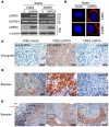
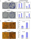
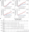
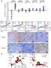
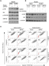
It has been recently reported that a side population of cells in nasopharyngeal carcinoma (NPC) displayed characteristics of stem-like cancer cells. However, the molecular mechanisms underlying the modulation of such stem-like cell populations in NPC remain unclear. Epstein-Barr virus was the first identified human tumor virus to be associated with various malignancies, most notably NPC. LMP2A, the Epstein-Barr virus encoded latent protein, has been reported to play roles in oncogenic processes. We report by immunostaining in our current study that LMP2A is overexpressed in 57.6% of the nasopharyngeal carcinoma tumors sampled and is mainly localized at the tumor invasive front. We found also in NPC cells that the exogenous expression of LMP2A greatly increases their invasive/migratory ability, induces epithelial-mesenchymal transition (EMT)-like cellular marker alterations, and stimulates stem cell side populations and the expression of stem cell markers. In addition, LMP2A enhances the transforming ability of cancer cells in both colony formation and soft agar assays, as well as the self-renewal ability of stem-like cancer cells in a spherical culture assay. Additionally, LMP2A increases the number of cancer initiating cells in a xenograft tumor formation assay. More importantly, the endogenous expression of LMP2A positively correlates with the expression of ABCG2 in NPC samples. Finally, we demonstrate that Akt inhibitor (V) greatly decreases the size of the stem cell side populations in LMP2A-expressing cells. Taken together, our data indicate that LMP2A induces EMT and stem-like cell self-renewal in NPC, suggesting a novel mechanism by which Epstein-Barr virus induces the initiation, metastasis and recurrence of NPC.
Conflict of interest statement
The authors have declared that no competing interests exist.
Figures


Similar articles
-
Epstein-Barr virus induction of the Hedgehog signalling pathway imposes a stem cell phenotype on human epithelial cells.J Pathol. 2013 Nov;231(3):367-77. doi: 10.1002/path.4245. J Pathol. 2013. PMID: 23934731
-
Regulation of DNA Damage Signaling and Cell Death Responses by Epstein-Barr Virus Latent Membrane Protein 1 (LMP1) and LMP2A in Nasopharyngeal Carcinoma Cells.J Virol. 2015 Aug;89(15):7612-24. doi: 10.1128/JVI.00958-15. Epub 2015 May 13. J Virol. 2015. PMID: 25972552 Free PMC article.
-
Cancer stem-like cell characteristics induced by EB virus-encoded LMP1 contribute to radioresistance in nasopharyngeal carcinoma by suppressing the p53-mediated apoptosis pathway.Cancer Lett. 2014 Mar 28;344(2):260-71. doi: 10.1016/j.canlet.2013.11.006. Epub 2013 Nov 19. Cancer Lett. 2014. PMID: 24262659
-
Novel roles and therapeutic targets of Epstein-Barr virus-encoded latent membrane protein 1-induced oncogenesis in nasopharyngeal carcinoma.Expert Rev Mol Med. 2015 Aug 18;17:e15. doi: 10.1017/erm.2015.13. Expert Rev Mol Med. 2015. PMID: 26282825 Review.
-
Modulation of the tumor microenvironment by Epstein-Barr virus latent membrane protein 1 in nasopharyngeal carcinoma.Cancer Sci. 2018 Feb;109(2):272-278. doi: 10.1111/cas.13473. Epub 2018 Jan 21. Cancer Sci. 2018. PMID: 29247573 Free PMC article. Review.
Cited by
-
The role of Epstein-Barr virus infection in the pathogenesis of nasopharyngeal carcinoma.Virol Sin. 2015 Apr;30(2):107-21. doi: 10.1007/s12250-015-3592-5. Epub 2015 Apr 21. Virol Sin. 2015. PMID: 25910483 Free PMC article. Review.
-
PXDN as a pan-cancer biomarker and promotes tumor progress via immune inhibition in nasopharyngeal carcinoma.Front Oncol. 2024 Sep 27;14:1463011. doi: 10.3389/fonc.2024.1463011. eCollection 2024. Front Oncol. 2024. PMID: 39399179 Free PMC article.
-
Epstein-Barr virus stably confers an invasive phenotype to epithelial cells through reprogramming of the WNT pathway.Oncotarget. 2018 Jan 2;9(12):10417-10435. doi: 10.18632/oncotarget.23824. eCollection 2018 Feb 13. Oncotarget. 2018. PMID: 29535816 Free PMC article.
-
Epstein-Barr virus-encoded latent membrane protein 2A promotes the epithelial-mesenchymal transition in nasopharyngeal carcinoma via metastatic tumor antigen 1 and mechanistic target of rapamycin signaling induction.J Virol. 2014 Oct;88(20):11872-85. doi: 10.1128/JVI.01867-14. Epub 2014 Aug 6. J Virol. 2014. PMID: 25100829 Free PMC article.
-
SYK interaction with ITGβ4 suppressed by Epstein-Barr virus LMP2A modulates migration and invasion of nasopharyngeal carcinoma cells.Oncogene. 2015 Aug 20;34(34):4491-9. doi: 10.1038/onc.2014.380. Epub 2014 Dec 22. Oncogene. 2015. PMID: 25531330
References
-
- Spano JP, Busson P, Atlan D, Bourhis J, Pignon JP, et al. Nasopharyngeal carcinomas: an update. Eur J Cancer. 2003;39:2121–2135. - PubMed
-
- Ong YK, Heng DM, Chung B, Leong SS, Wee J, et al. Design of a prognostic index score for metastatic nasopharyngeal carcinoma. Eur J Cancer. 2003;39:1535–1541. - PubMed
-
- Lee AW, Poon YF, Foo W, Law SC, Cheung FK, et al. Retrospective analysis of 5037 patients with nasopharyngeal carcinoma treated during 1976-1985: overall survival and patterns of failure. Int J Radiat Oncol Biol Phys. 1992;23:261–270. - PubMed
-
- Thiery JP, Sleeman JP. Complex networks orchestrate epithelial-mesenchymal transitions. Nat Rev Mol Cell Biol. 2006;7:131–142. - PubMed
Publication types
MeSH terms
Substances
LinkOut - more resources
Full Text Sources
Molecular Biology Databases

