. 2010 Jun 1;8(6):e1000386.
doi: 10.1371/journal.pbio.1000386.

A feed-forward circuit linking wingless, fat-dachsous signaling, and the warts-hippo pathway to Drosophila wing growth

Affiliations

A feed-forward circuit linking wingless, fat-dachsous signaling, and the warts-hippo pathway to Drosophila wing growth

Myriam Zecca et al. PLoS Biol. .

Abstract

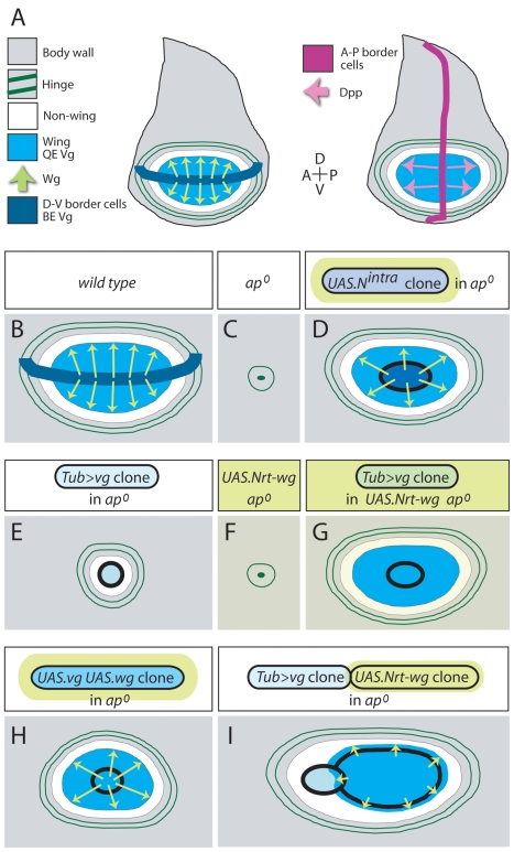
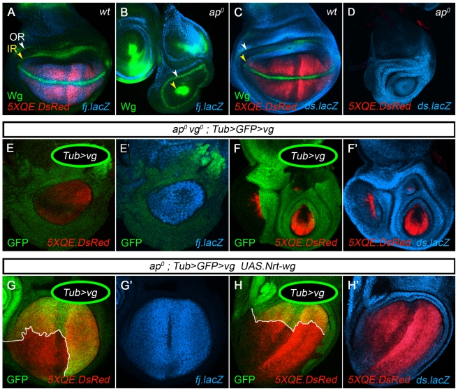
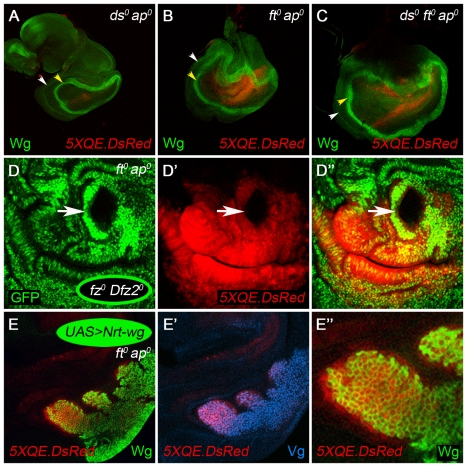
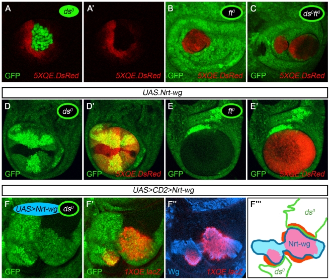
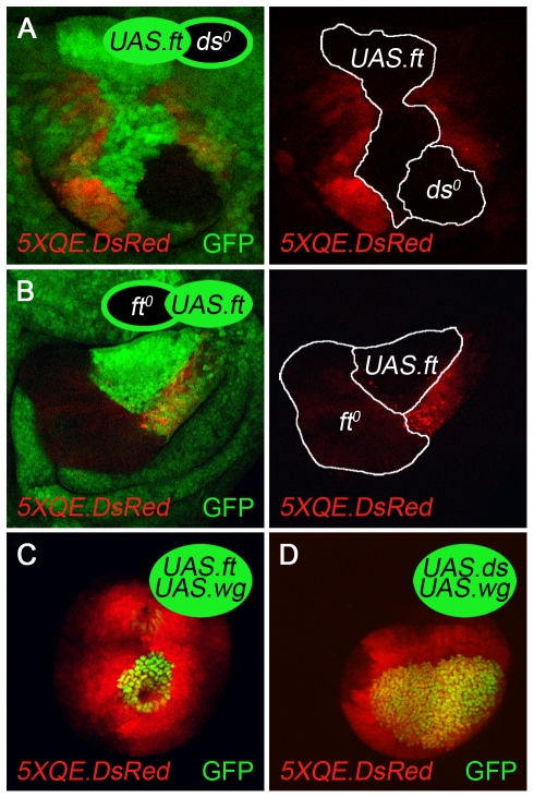
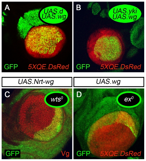
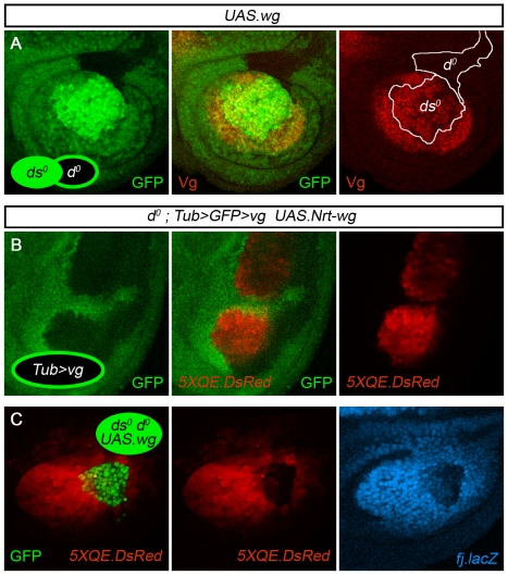
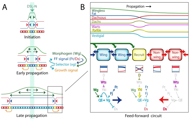
During development, the Drosophila wing primordium undergoes a dramatic increase in cell number and mass under the control of the long-range morphogens Wingless (Wg, a Wnt) and Decapentaplegic (Dpp, a BMP). This process depends in part on the capacity of wing cells to recruit neighboring, non-wing cells into the wing primordium. Wing cells are defined by activity of the selector gene vestigial (vg) and recruitment entails the production of a vg-dependent "feed-forward signal" that acts together with morphogen to induce vg expression in neighboring non-wing cells. Here, we identify the protocadherins Fat (Ft) and Dachsous (Ds), the Warts-Hippo tumor suppressor pathway, and the transcriptional co-activator Yorkie (Yki, a YES associated protein, or YAP) as components of the feed-forward signaling mechanism, and we show how this mechanism promotes wing growth in response to Wg. We find that vg generates the feed-forward signal by creating a steep differential in Ft-Ds signaling between wing and non-wing cells. This differential down-regulates Warts-Hippo pathway activity in non-wing cells, leading to a burst of Yki activity and the induction of vg in response to Wg. We posit that Wg propels wing growth at least in part by fueling a wave front of Ft-Ds signaling that propagates vg expression from one cell to the next.


Conflict of interest statement

The authors have declared that no competing interests exist.

Figures

Figure 1
Figure 1. Feed-forward signaling: context and criteria.
(A) Context. Two diagrams of the mature wing imaginal disc are shown, depicting control of wing growth by Wg (left) and Dpp (right) and keys for the relevant primordia, signals, and gene expression domains. Early in larval life, the wing disc is subdivided into distal (prospective wing; turquoise/white) and proximal (prospective hinge and body wall; grey) domains. Feed-forward (FF) signaling operates only in the distal domain, to induce non-wing cells (white) to enter the wing primordium (turquoise). Both domains are further subdivided into D and V compartments by activity of the selector gene ap in the D compartment (not depicted). DSL-Notch signaling across the D-V compartment boundary defines a population of specialized border cells (dark blue) that express wg and vg, the latter mediated by the vg Boundary enhancer (BE). The wing disc is also divided into anterior (A) and posterior (P) compartments, with A cells just anterior to the A-P boundary secreting Dpp (for simplicity only shown in A). Following the D-V segregation, vg expressing wing cells send a short-range feed-forward (FF) signal (not depicted) that acts together with Wg and Dpp to activate Quadrant enhancer (QE) dependent vg expression (turquoise) in abutting non-wing cells; newly recruited wing cells serve as a source for new FF signal, propagating recruitment of neighboring non-wing cells into the wing primordium in response to Wg and Dpp (see Figure 8A). Wg and Dpp are also required (i) to maintain QE-dependent vg expression in cells once they are recruited into the wing primordium, (ii) to sustain the survival and growth of wing cells, so defined, and (iii) to act indirectly, through the action of Vg, to produce an additional signal that induces proliferation of surrounding non-wing cells for recruitment into the growing wing primordium ,. The hinge primordium, which encircles the prospective wing, contains two concentric rings of wg expressing cells (dark green) that serve as landmarks as well as potential sources for cryptic Wg signal in apo discs. (B–I) Criteria. FF signaling is monitored by assaying QE-dependent gene expression. (B) wild type. Here, as in the remaining panels, the genotype is indicated above and the QE response below for each of several experimental paradigms used to define the FF signal ,. Wg signal is depicted by Chartreuse arrows or wash. QE activity and formation of wing tissue (turquoise) indicates a positive response. (C) The apo condition serves as the ground state for assaying FF signaling. In the absence of ap, no D-V segregation occurs, no D-V border cells are specified and the nascent wing primordium ceases to express vg, yielding a population of “non-wing” cells that either die or sort out during subsequent development, unless they are induced to activate QE-dependent vg expression in response to Wg and the FF signal generated by an experimental manipulation (Dpp is provided, independently, by A-P border cells). As diagrammed, mature apo discs lack wing (turquoise) and non-wing (white) territories, as well as the distal portion of the hinge primordium, reducing the inner ring of Wg expression to a small patch, encircled by a rudimentary outer ring. (D) Cells that express constitutively active forms of Notch in apo discs (e.g., UAS.Nintra clones) behave like ectopic D-V border cells. They express wg and vg, induce neighboring non-wing cells to activate QE-dependent vg expression, and recruit surrounding cells to join a rapidly expanding wing primordium. (E,F) Providing only Vg expressing cells [e.g., Tub>vg clones; (E)] or only ectopic Wg signal [uniform expression of Neurotactin-Wg (UAS.Nrt-Wg), a membrane tethered form of Wg] fails to induce QE activity, except within Tub>vg expressing cells, where the combination of cryptic Wg input and exogenous Vg activity weakly activates the QE cell-autonomously (E, light turquoise wash). (G–I) Generating Vg expressing cells in the presence of Wg signal, whether in the form of ubiquitous Nrt-Wg expression (G), co-expression of ectopic Wg (H), or abutting clones of Nrt-Wg expressing cells (I), induces long-range propagation of QE-dependent vg expression and rescue of wing tissue. Note that in the last condition (I), FF signaling can propagate throughout the Nrt-Wg clone and extend to abutting wild type cells (which receive the Nrt-Wg signal) but does not go further owing to inadequate Wg signal in the surround.
Figure 2
Figure 2. Vestigial activates four-jointed and represses dachsous.
(A–D) fj-lacZ, ds-lacZ, and 5XQE.DsRed reporter expression in mature wild type and apo discs counter-stained for Wg (A–C, only); note that 5XQE.DsRed expression is reduced in the vicinity of the A-P compartment boundary, as also apparent in (G,H). In wild type discs (A,C), fj-lacZ and 5XQE.DsRed are co-expressed in the wing pouch in a domain complementary to that of ds-lacZ (the inner (IR) and outer (OR) rings of Wg in the hinge primordium are indicated by yellow and white arrow heads). In apo discs (B,D), the wing pouch is absent, as indicated by the collapse of the IR to a small circular patch surrounded by the OR: ds-lacZ is expressed uniformly in place of fj-lacZ and 5XQE.DsRed in the territory encircled by the OR. (E,F). fj-lacZ, ds-lacZ, and 5XQE.DsRed reporter expression in apo vgo discs that contain clones of Tub>vg cells (marked by the absence of GFP). Clones located in the prospective wing domain develop cell-autonomously as wing tissue and express fj-lacZ and 5XQE.DsRed instead of ds-lacZ. (G,H) Clones of Tub>vg cells in UAS.Nrt-wg expressing apo discs (as in Figure 1G). Clones (outlined in white, marked by the absence of GFP) induce the long-range propagation of QE-dependent vg expression, as visualized by the domain of 5XQE.DsRed expression. Recruitment into wing tissue correlates with the up-regulation of fj-lacZ expression and the down-regulation of ds-lacZ expression. Here, and in subsequent figures, genotypes, clone markers, and antibody stains are indicated on the panels, coded by color (clones marked by the absence of GFP are shown as open circles with green borders; those marked positively are shown as filled circles), or in boxes above the panels (in all cases in which a UAS transgene is indicated in a box, its expression is driven by a Gal4 driver that is uniformly active in the prospective wing territory; white/turquoise domain as in Figure 1A, 1B; see Materials and Methods for exact genotypes).
Figure 3
Figure 3. Fat and Dachsous are required to block Quadrant enhancer activity in the absence of feed-forward signal.
(A–C) Removal of either, or both, Ft and Ds causes constitutive, low-level QE activity (monitored by 5XQE.DsRed expression) in apo discs. apo discs that are dso, fto, or dso fto form wing pouches that express the 5XQE.DsRed reporter and are encircled by the Wg IR and OR, in contrast to single mutant apo discs (Figure 2B). Note that the level of 5XQE.DsRed expression is very low, especially in the dso apo disc, consistent with the presence of only cryptic levels of Wg; note also that some DsRed expression within the rescued pouch appears outside of the Wg IR because it is in a fold, underneath. (D) The 5XQE.DsRed response observed in fto apo discs depends on Wg input. 5XQE.DsRed expression is lost in clones of fzo Dfz2o cells in the wing pouch of fto apo discs (a single fzo Dfz2o clone is indicated by an arrow). (E) Clones of UAS.Nrt-wg cells induce normal, peak expression of both the 5XQE.DsRed reporter and endogenous Vg within the clone and in adjacent cells (the low levels of 5XQE.DsRed and Vg expression in surrounding cells can only be detected, as in A–D, using more intense laser illumination).
Figure 4
Figure 4. Fat is required in vestigial expressing cells to send feed-forward signal.
(A–C) Clones of dso, fto, and dso fto cells in apo discs. The dso clone (A) is marked positively by the expression of GFP to allow the non-autonomous induction of 5XQE.DsRed expression to be clearly distinguished from the clone. Conversely, fto and dso fto clones (B,C) are marked negatively, by the absence of GFP, to visualize the strictly cell-autonomous expression of the 5XQE.DsRed transgene. 5XQE.DsRed is expressed only at cryptic low levels within dso, fto, and dso fto clones (as in entirely dso apo, fto apo, and dso fto apo discs; Figure 3A–C) and is not detectable in (A) at the level of laser illumination used to generate this image. However, dso clones induce surrounding, wild type cells to express much higher levels of 5XQE.DsRed expression, in contrast to fto and dso fto clones, indicating that they generate ectopic FF signal. We infer that the absence of Ds activity in the dso cells constitutively activates the FF signal transduction pathway but only at a low level relative to the peak response of surrounding, wild type cells to ectopic FF signal sent by the clone. (D,E) Clones of dso and fto cells in apo discs that express UAS.Nrt.wg uniformly under Gal4 control (“UAS.Nrt-wg” discs in all subsequent panels; the non-autonomous induction of 5XQE.DsRed expression appears as yellow in D'). dso clones activate 5XQE.DsRed expression cell-autonomously and serve as a potent source of FF signal, inducing surrounding cells to express the 5XQE.DsRed reporter and join a growing wing primordium. Conversely, most fto clones show only a strictly cell-autonomous response (exceptions appear to be associated with ectopic FF signal generated by sibling ft+/ft+ clones, as documented in Figure S2). (F) An apo disc containing abutting dso and UAS.Nrt-wg clones marked, respectively, by the absence of GFP and the expression of Nrt-Wg (F''' depicts the experiment in cartoon form). The dso clones behave like Tub>vg clones (Figure 1I): they induce high levels of QE-dependent vg expression (monitored by both 1XQE.lacZ and endogenous Vg expression) in the abutting Nrt-Wg cells within the prospective wing domain. Moreover, QE activation propagates over many cell diameters within the Nrt-Wg clone and extends to adjacent cells across the clone border. Finally, the QE response is also up-regulated in dso cells that abut the Nrt-Wg clone, in response to the tethered Wg signal.
Figure 5
Figure 5. Generation and transduction of feed-forward signal by Fat and Dachsous in apo discs.
(A) Abutting, sibling clones of UAS.ft and dso cells marked, respectively, by high (2×) or no (0×) GFP expression in a background of moderate (1×) GFP expressing cells, and outlined in white. The UAS.ft clone has induced 5XQE.DsRed expression in neighboring wild type cells but not in the abutting dso cells. As noted in the legend to Figure 4A, the loss of ds is associated with the cell-autonomous activity of the 5XQE-DsRed reporter but only at cryptic, low level relative to the response induced in wild type cells by receipt of FF signal (and hence not detected at the level of laser illumination used in this image). (B) Abutting, sibling clones of UAS.ft and fto cells (marked as in A). The result is the same: like dso cells, the fto cells are refractory to induction of the 5XQE-DsRed transgene by abutting UAS.ft cells, in contrast to neighboring wild type cells. Similarly, as in the case of dso clones, 5XQE.DsRed transgene is expressed constitutively, but only at cryptic low level, in fto clones (as in Figure 4B) and is not readily detectable in this image. (C,D) UAS.ft UAS.wg (C) and UAS.ds UAS.wg (D) clones: The 5XQE.DsRed transgene is strongly expressed both within, and in a halo around, each clone.
Figure 6
Figure 6. Reducing or bypassing Warts-Hippo activity ectopically activates Quadrant enhancer-dependent vestigial expression.
(A,B) Clones of UAS.d (A) and UAS.yki (B) cells that co-express UAS.wg in apo discs. Both clones activate QE dependent gene expression cell-autonomously as monitored by 5XQE.DsRed expression. Both have also induced 5XQE.DsRed expression in neighboring cells encircling the clone. (C,D) Clones of wtso (C) and exo (D) cells in UAS.Nrt-wg apo discs. Both clones activate QE-dependent gene expression cell-autonomously and have also induced QE activity in neighboring cells (monitored by Vg in C, and 5XQE.DsRed expression in D).
Figure 7
Figure 7. Dachs is required to receive, but not to send, feed-forward signal.
(A) Sibling clones of dso and do cells in an UAS.wg apo disc (clones marked by 2× and 0× GFP expression, respectively, as in Figure 5A, and outlined in white). The dso clone expresses Vg and has induced Vg expression in abutting wild type cells but not in abutting do cells. As a consequence, the latter are unable to contribute to the rescued wing primordium. This result contrasts with the behavior of wild type clones that are generated as siblings of dso clones: as shown in Figure 4D, cells within such wild type clones can respond by activating the QE and joining the wing primordium. (B) Two clones of Tub>vg cells in an UAS.Nrt-wg do apo disc. Both clones express the 5XQE-DsRed reporter cell-autonomously and have induced a few adjacent cells to do the same, in marked contrast to the long-range propagation of QE-dependent Vg expression associated with Tub>vg clones generated in UAS.Nrt-wg apo discs that retain wild type d function (Figures 1G, 2G). (C) A UAS.wg dso do clone in an apo disc. The clone has induced the long-range propagation of 5XQE-DsRed and fj-lacZ expression in surrounding cells, but cells within the clone have failed to respond, or express only low levels of both reporters, indicating that they can send, but not receive, the FF signal.
Figure 8
Figure 8. The vestigial feed-forward circuit, and the control of wing growth by morphogen.
(A) A model for the control of wing growth by Wg. Initiation (top): the main phase of wing growth begins with segregation of the wing disc into D-V compartments and the induction of specialized border cells (dark blue) by DSL-Notch signaling (mint green): Notch activity drives expression of both the morphogen Wg (dark green) as well as Boundary enhancer (BE) dependent expression of the wing selector gene vg. As detailed in (B), Vg activity up-regulates Ft signaling (blue) at the expense of Ds signaling (red) to generate the feed-forward (FF) signal. The FF signal then acts together with Wg to induce Quadrant enhancer (QE) expression of vg in non-wing cells (red), initiating a stable circuit of Wg-dependent vg expression that recruits the responding non-wing cell (yellow) into the wing primordium. Early propagation (middle): Vg activity in newly recruited wing cells (turquoise) generates new FF signal, which acts together with Wg secreted by border cells to induce QE-dependent vg expression in neighboring non-wing cells. It also leads to the production of an additional “growth” signal (orange arrows) that promotes proliferation of the surrounding population of non-wing cells from which new wing cells will be recruited. As shown to the right, Vg activity and FF signaling comprise an auto-regulatory cycle driven by Wg. Each turn of the cycle corresponds to the recruitment of a non-wing cell into the wing primordium and generates a new, non-wing cell for subsequent recruitment. Late propagation (bottom): The wing primordium increases in size, propelled by propagation of the FF recruitment cycle and proliferation of cells within and around the primordium, both fueled by Wg as it spreads from D-V border cells. FF forward propagation also depends on Dpp spreading from border cells along the A-P compartment boundary, which acts together with Wg to promote the outward growth of the wing primordium from the intersection between the D-V and A-P compartment boundaries (not depicted; Figure 1A). (B) The feed-forward circuit. Top: the signaling activities of Wg, Ft, and Ds as well as the transducing activities of D, Wts, and Yki are shown relative to vg transcription, and the recruitment of non-wing cells into the wing primordium (recruitment propagates from left to right, coloring as in A). Away from the recruitment interface, Ft and Ds signaling activities are weakly graded or flat, D and Yki activities are low, and Wts activity is high. At the recruitment interface, Ft and Ds signaling activities are steeply graded and opposite, generating a transient pulse in D activity, a dip in Wts activity, and a burst of Yki activity. Middle: Wg, Ft, and Ds signals are shown as green, blue, and red arrows. Only the cell undergoing recruitment (yellow) receives both Wg as well as steep and opposing Ft and Ds signals. Bottom: the regulatory circuits underlying the wing (vg ON; left) and non-wing (vg OFF; right) states as well as the transition that occurs during recruitment (vg OFF to vg ON; middle) are diagrammed relative to the landscapes of Wg, Ft, and Ds signaling upon which they depend. In wing cells, Wg input acts together with Vg to drive a positive auto-regulatory circuit of vg expression mediated by the Quadrant Enhancer (QE), and Vg up-regulates the expression of Fj while repressing that of Ds to enhance Ft signaling at the expense of Ds signaling (blue arrow). In non-wing cells, both the absence of Vg as well as the low level of nuclear Yki lead, by default, to low levels of Fj and high levels of Ds, enhancing Ds signaling at the expense of Ft signaling (red arrow). The box underneath each cell depicts the level and asymmetry of Ft and Ds inputs received from abutting cells on either side. Relatively uniform inputs (depicted by parallel lines in the boxes under wing and non-wing cells) cause modest, or no, polarization of the transducing activities of both proteins within each cell, suppressing the capacity of D to inhibit Wts activity and elevate nuclear import of Yki. Conversely, steep and opposite inputs (depicted by crossing lines in the box beneath the cell undergoing recruitment) cause a strong polarization, allowing D to inhibit Wts activity and induce a burst of Yki nuclear activity. Both Yki and Vg activate vg transcription via the QE by functioning as transcriptional co-activators for the same DNA binding protein, Sd (not depicted). Hence, as the level of Vg rises in cells undergoing recruitment, Vg can substitute for Yki to generate a stable circuit of Wg-dependent Vg auto-regulation that no longer requires Yki or FF input. Note that the depictions of vg expression, as well as of Ft and Ds signaling, as uniform away from the recruitment interface are simplifications. Instead, vg expression is weakly graded within the wing primordium in response to graded Wg signal, and the complementary patterns of fj and ds, upon which the signaling activities of Ft and Ds depend, are similarly graded. The resulting shallow differentials of Ft and Ds signaling may suffice to polarize cells in the plane of the epithelium (PCP). Nevertheless, the expression profiles of all three genes show a dramatic increase in steepness at the periphery of the wing primordium, and it is the resulting steepness in opposing Ds and Ft signals that we posit is essential to induce the burst of Yki nuclear activity upon which recruitment depends.

Comment in

Similar articles

Cited by

References

    1. Zecca M, Basler K, Struhl G. Sequential organizing activities of engrailed, hedgehog and decapentaplegic in the Drosophila wing. Development. 1995;121:2265–2278. - PubMed
    1. Nellen D, Burke R, Struhl G, Basler K. Direct and long-range action of a DPP morphogen gradient. Cell. 1996;85:357–368. - PubMed
    1. Lecuit T, Brook W. J, Ng M, Calleja M, Sun H, et al. Two distinct mechanisms for long-range patterning by Decapentaplegic in the Drosophila wing. Nature. 1996;381:387–393. - PubMed
    1. Zecca M, Basler K, Struhl G. Direct and long-range action of a wingless morphogen gradient. Cell. 1996;87:833–844. - PubMed
    1. Neumann C. J, Cohen S. M. Long-range action of Wingless organizes the dorsal-ventral axis of the Drosophila wing. Development. 1997;124:871–880. - PubMed

Publication types

MeSH terms

LinkOut - more resources