. 2010 Jun 1;8(6):e1000388.
doi: 10.1371/journal.pbio.1000388.

Defensin-like ZmES4 mediates pollen tube burst in maize via opening of the potassium channel KZM1

Affiliations

Defensin-like ZmES4 mediates pollen tube burst in maize via opening of the potassium channel KZM1

Suseno Amien et al. PLoS Biol. .

Abstract

In contrast to animals and lower plant species, sperm cells of flowering plants are non-motile and are transported to the female gametes via the pollen tube, i.e. the male gametophyte. Upon arrival at the female gametophyte two sperm cells are discharged into the receptive synergid cell to execute double fertilization. The first players involved in inter-gametophyte signaling to attract pollen tubes and to arrest their growth have been recently identified. In contrast the physiological mechanisms leading to pollen tube burst and thus sperm discharge remained elusive. Here, we describe the role of polymorphic defensin-like cysteine-rich proteins ZmES1-4 (Zea mays embryo sac) from maize, leading to pollen tube growth arrest, burst, and explosive sperm release. ZmES1-4 genes are exclusively expressed in the cells of the female gametophyte. ZmES4-GFP fusion proteins accumulate in vesicles at the secretory zone of mature synergid cells and are released during the fertilization process. Using RNAi knock-down and synthetic ZmES4 proteins, we found that ZmES4 induces pollen tube burst in a species-preferential manner. Pollen tube plasma membrane depolarization, which occurs immediately after ZmES4 application, as well as channel blocker experiments point to a role of K(+)-influx in the pollen tube rupture mechanism. Finally, we discovered the intrinsic rectifying K(+) channel KZM1 as a direct target of ZmES4. Following ZmES4 application, KZM1 opens at physiological membrane potentials and closes after wash-out. In conclusion, we suggest that vesicles containing ZmES4 are released from the synergid cells upon male-female gametophyte signaling. Subsequent interaction between ZmES4 and KZM1 results in channel opening and K(+) influx. We further suggest that K(+) influx leads to water uptake and culminates in osmotic tube burst. The species-preferential activity of polymorphic ZmES4 indicates that the mechanism described represents a pre-zygotic hybridization barrier and may be a component of reproductive isolation in plants.


Conflict of interest statement

The authors have declared that no competing interests exist.

Figures
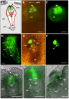
Figure 1
Figure 1. ZmES4 is predominately localized to the secretory zone of the mature maize egg apparatus before fertilization.
(A) Diagram of the female gametophyte (embryo sac) of maize embedded in the maternal tissues of the ovule. Outer integument is black, inner integument dark grey, and nucellus light grey. The egg cell (blue) is hidden behind the two synergids and the filiform apparatus is indicated with a white arrowhead. (B) Merged bright-field and UV micrograph showing a slanting oversight of the micropylar region. The inner integument has been removed. ZmES4-GFP fusion protein under control of the endogenous promoter is localized to the synergid cells with strongest signals around the filiform apparatus (arrowhead). The arrow points towards the five to six micropylar nucellus cell stacks enclosing the egg apparatus. (C) Enhanced UV image of (B) to show strongest GFP signals in the secretory zone of the two synergid cells (arrows). Vesicles are visible in the synergid extensions within the filiform apparatus (arrow head). Weaker signals are visible in the central cell. (D) CLSM image stack of 30 1 µm sections of a young mature embryo sac. The ZmES4-GFP fusion protein is visible in cytoplasmic strands probably resembling the ER and vesicles of the egg apparatus (egg cell and synergids) and central cell but not in the antipodals. Highest signals are visible in the egg apparatus. (E) Merged bright-field and UV micrograph showing a section along the embryo sac 15 h after in vitro pollination with a view from the inside of the ovule. The surface of micropylar nucellus cell stacks is indicated by asterisks. (F) UV image of (E) to show that GFP signals are no longer visible in the cell wall of the egg apparatus, in the receptive and degenerated synergid cell, as well as in the central cell. Strongest signal is detectable in the persisting synergid cell and weaker signals around the nucleus of the zygote. 24 h after in vitro pollination, signals were no longer detectable (unpublished data). (G–I) Longitudinal sections of a marker line displaying GFP only in one of the two synergid cells. View from the inside of the ovule towards the female gametophyte. (G) A young embryo sac shortly after cellularization displays GFP signals exclusively in the synergid cell. (H) A mature embryo sac of the same line shows strong GFP signals around the nucleus (asterisk) and in the secretory zone (arrowhead) of one synergid cell. (I) The GFP expressing synergid cell is the exclusive recipient of pollen tubes, indicated by the loss of GFP signal. AP, antipodals; CC, central cell; EC, egg cell; II, inner integument; mNU, micropylar nucellus; NU, nucellus; OI, outer integument; SY, synergid; dSY, degenerated synergid; pSY, persisting synergid; PN, polar nuclei of the central cell; PT, pollen tube; Z, zygote; nZ, nucleus of zygote. Scale bars are 50 µm.
Figure 2
Figure 2. Pollen tube arrest and burst is impaired in transgenic lines with reduced ZmES activity.
(A) In order to study the function of ZmES genes, ZmES4-RNAi mutants were generated to down-regulate the whole ZmES gene-family simultaneously by RNA silencing. Only about half of self-pollinated ovules of ZmES4-RNAi lines developed into kernels (left). Pollination of wt (A188) ovules using pollen from ZmES4-RNAi plants led to full seed set (right) and about 43% transmission of a single copy RNAi construct in progeny plants (unpublished data). (B–G) ACTp:GUS maize pollen was used to monitor pollen tube growth and fertilization rates inside maize ovaries between 27–32 hap (B–E) and 72 hap (F and G), respectively. (B) One pollen tube (arrow) has penetrated the egg apparatus of a wt ovule. Blue staining of both egg apparatus and central cell indicates successful double fertilization. A second pollen tube (arrowhead) is no longer directed towards the fertilized egg apparatus. (C) An ACTp:GUS maize pollen tube has penetrated the nucellar tissue (arrow) and female gametophyte (arrow head) of an ZmES4-RNAi ovule, but GUS signals are neither detectable in the synergid nor in the central cell. (D) A close-up of (C) shows that the pollen tube grew into the cell wall material of the filiform apparatus between the two synergids (arrowhead). GUS activity is visible inside the female gametophyte in the region between egg and central cell indicating pollen tube growth arrest. (E) Another example of an ZmES4-RNAi ovule containing a pollen tube inside the egg apparatus region lacking both growth arrest and burst. (F) A wt ovule shows paternal GUS-activity in both pro-embryo and developing endosperm. The endosperm has already enlarged and contains a large number of nuclei (not visible). (G) The nucellus tissue (arrow) of an ZmES4-RNAi ovule was penetrated by a pollen tube, which grew around the egg apparatus (arrowhead) without penetration. The synergid cells have already started to degenerate at this stage. (H) Fertilization rates of both egg and central cell are significantly impaired in ZmES4-RNAi ovules. On average 88% wt ovules are fertilized (black columns), compared to 60% fertilized ZmES4-RNAi ovules (grey columns). A difference between self-pollinated ZmES4-RNAi ovules as well as ovules from back-crossed (BC) plants was not detectable neither 1 nor 2 d after fertilization (27–32 and 53–55 hap, respectively). The percentage of unfertilized ovules for both wt and ES4-RNAi lines are represented by white columns. A detailed list of sectioned and analyzed ovaries is attached as Table S1. AP, antipodals; CC, central cell; EA, egg apparatus; EN, endosperm; FA, filiform apparatus; hap, hours after in vitro pollination; mNU, micropylar nucellus; PE, pro-embryo; SY, synergid. Scale bars are 50 µm.
Figure 3
Figure 3. Mature defensin-like ZmES4 induces pollen tube tip burst after depolarization of the pollen tube membrane.
In order to investigate the role of ZmES4 activity on living pollen tubes, an in vitro pollen tube growth bioassay using various plant species was established. (A) A pollen grain of maize (inbred A188) was germinated for 15 min in liquid medium before addition of 30 µM RsAFP2 in PGM. Growth behavior was further observed for up to 120 min (here after 15 min incubation) displaying normal tube growth. (B) Maize pollen grains were germinated for 15 min before addition of PGM containing 30 µM ZmES4. Pollen tubes started to burst within seconds after ZmES4 application and tube content was released explosively at the tip (arrow). (C) Pollen grains of Tripsacum dactyloides were germinated for 15 min before PGM containing 30 µM ZmES4 was added. 15 min after ZmES4 application most pollen tubes were still intact and grew further until rupture occurred on average around 32 min after incubation. (D) Diagram showing average time points when 80% of germinated pollen tubes of maize (Zea mays) and T. dactyloides were ruptured after ZmES4 protein application. Growth of pollen tubes of lily (H. fulva) and various tobacco species (N. spec.) observed for more than 60 min was not affected by ZmES4 application. CsCl2 application significantly delayed ZmES4 activity. Standard deviations are indicated. (E) Micro-electrode impalement of Zea mays pollen tubes for membrane potential recordings. Pollen grains of maize were germinated on solid medium and impaled right below the pollen tube tip. Control application of CRPs such as trypsin inhibitor from soybean or RsAFP2 in PGM did not affect the pollen tube membrane potential, while the application of ZmES4 in PGM evoked a transient depolarization of the membrane potential followed by pollen tube tip burst (arrowhead). A representative recording is shown displaying tube burst 2 min after ZmES4 application. Bars: 50 µm.
Figure 4
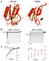
Figure 4. ZmES4 opens the pollen tube expressed potassium channel KZM1.
(A) X-ray structure of Sahara scorpion (Androctonus australis) neurotoxin II (AaToxII: PDB code 1PTX; left) and predicted 3D-structure of ZmES1 of maize (right). The ZmES1 3D-structure was generated using the program MODELLER based on various defensin NMR structures. The structure is almost identical to that of ZmES4 (Figure S3) and displays the predicted mature protein. All 3D-structures are composed of a α-helix as well as a three-stranded anti-parallel β-sheet. Four disulfide bonds between C1 and C8 are painted in green. The arrow points towards the loop between C5 and C6 of ZmES1, which may be responsible for ZmES specificity. (B) ZmES4 modulates the activity of the potassium channel KZM1. Electrophysiological recordings of ZmES4 action on KZM1 expressed in Xenopus oocytes. Typical voltage dependent inward rectifying potassium currents of KZM1 in bath medium with and without control substances such as trypsin inhibitor from soybean or antifungal protein AFP2 from Raphanus sativus (left). Loss of voltage dependent gating of KZM1 upon addition of ZmES4 (150 µg/ml) in the bath medium (right). (C) Current voltage relation of KZM1 steady state currents in response to 150 µg/ml ZmES4. Recordings were performed in a bath solution containing 100 mM KCl. Currents were evoked upon voltage jumps in the range of +50 to −180 mV. Data points of four independent experiments were normalized to the value at −160 mV in control solution. The graph on the right depicts a magnification of the data shown on the left in the voltage range between −60 and −100 mV. Due to the loss of voltage dependence in the presence of ZmES4, KZM1 mediates K+ currents at all tested membrane potentials. Error bars indicate the standard deviation.

Similar articles

Cited by

References

    1. Bell C. D, Soltis D. E, Soltis P. S. The age of the angiosperms: a molecular timescale without a clock. Evolution. 2005;59:1245–1258. - PubMed
    1. Punwani J. A, Drews G. N. Development and function of the synergid cell. Sex Plant Reprod. 2008;21:7–15.
    1. Dresselhaus T. Cell-cell communication during double fertilization. Curr Opin Plant Biol. 2006;9:41–47. - PubMed
    1. Strasburger E. Neuere untersuchungen über den befruchtungsvorgang bei den phanerogamen als grundlage für eine theorie der zeugung. Jena, Gustav Fischer 1884
    1. Escobar-Restrepo J. M, Huck N, Kessler S, Gagliardini V, Gheyselinck J, et al. The FERONIA receptor-like kinase mediates male-female interactions during pollen tube reception. Science. 2007;317:656–660. - PubMed

Publication types