

Persistent left superior vena cava with absent right superior vena cava: a case report and review of the literature
- PMID: 20532458
- PMCID: PMC5592325
Persistent left superior vena cava with absent right superior vena cava: a case report and review of the literature
Abstract
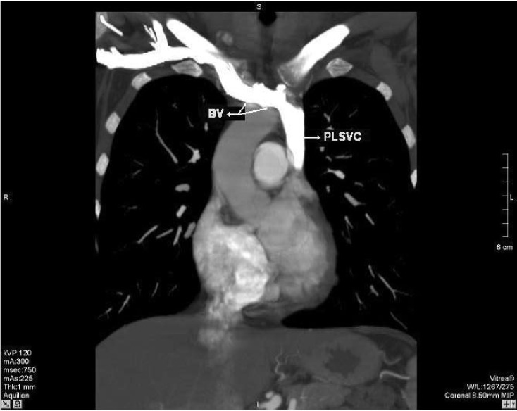
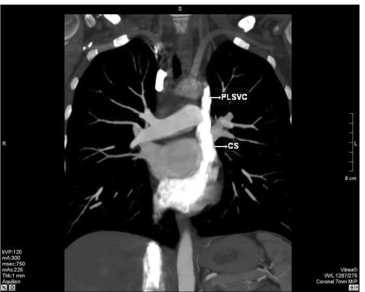
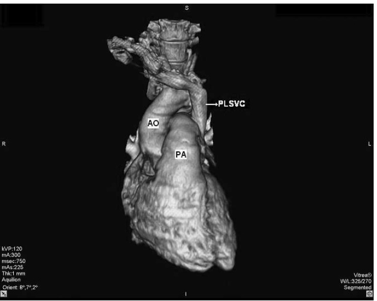
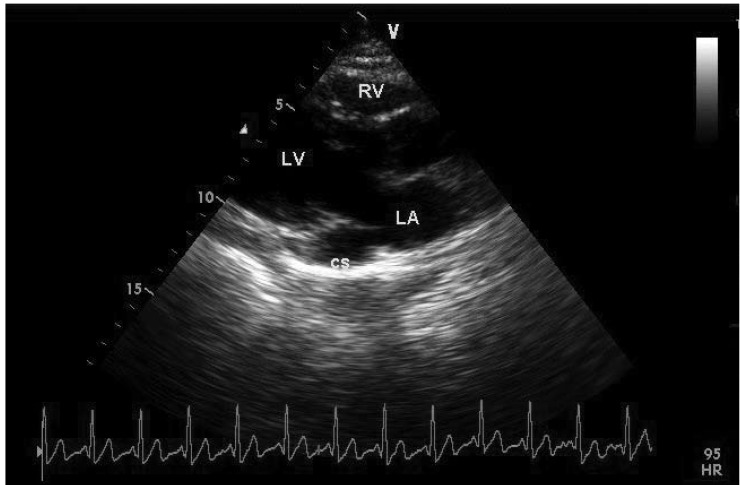
We report on a rare case of persistent left superior vena cava (PLSVC) with absent right superior vena cava (RSVC), an anomaly that is also known as isolated PLSVC. This venous malformation was identified incidentally in a 30-year-old woman during thoracic multi-detector computed tomography (MDCT), which was performed with the suspicion of intra-thoracic malignancy. On thoracic MDCT, the RSVC was absent. A bridging vein drained the right jugular and right subclavian veins and joined the left brachiocephalic vein in order to form the PLSVC, which descended on the left side of the mediastinum and drained into the right atrium (RA) via a dilated coronary sinus (CS). The patient was referred to the cardiology department for further evaluation. Transthoracic echocardiography revealed a dilated CS, and agitated saline injected from the left or right arms revealed opacification of the CS before the RA. The patient had no additional cardiac abnormality. Isolated PLSVC is usually asymptomatic but it can pose difficulties with central venous access, pacemaker implantation and cardiothoracic surgery. This condition is also associated with an increased incidence of congenital heart disease, arrhythmias and conduction disturbances. A wide spectrum of clinicians should be aware of this anomaly, its variations and possible complications.
Figures
References
-
- Danielpour PJ, Aalberg JK, El-Ramey M, Sivina M, Wodnicki H. Persistent left superior vena cava:an incidental finding during central venous catheterisation – a case report. Vasc Endovascular Surg. 2005;39:109–111. - PubMed
-
- Bartram U, van Praagh S, Levine JC, Hines M, Bensky AS, van Praagh R. Absent right superior vena cava in visceroatrial situs solitus. Am J Cardiol. 1997;80:175–183. - PubMed
-
- Fischer DR, Zuberbuhler JR. Paediatric Cardiology. New York: churchill Livingstone: 2002. Anomalous systemic venous return. In: Anderson RH, Baker E, Mccartney RF, eds. pp. 851–865.
-
- Shyamkumar NK, Brown R. Double superior vena cava with a persistent left superior vena cava: an incidental finding during peripherally inserted central catheter placement. Australas Radiol. 2007;51(Suppl B):257–259. - PubMed
Publication types
MeSH terms
LinkOut - more resources
Full Text Sources
