

An automated neural-fuzzy approach to malignant tumor localization in 2D ultrasonic images of the prostate
- PMID: 20532587
- PMCID: PMC3092054
- DOI: 10.1007/s10278-010-9301-x
An automated neural-fuzzy approach to malignant tumor localization in 2D ultrasonic images of the prostate
Abstract
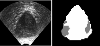
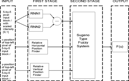
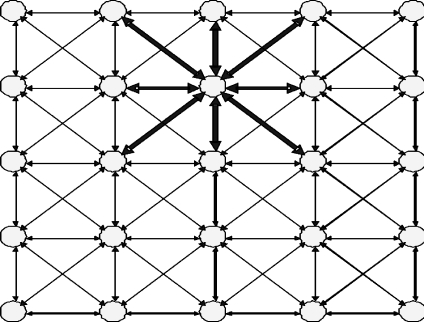
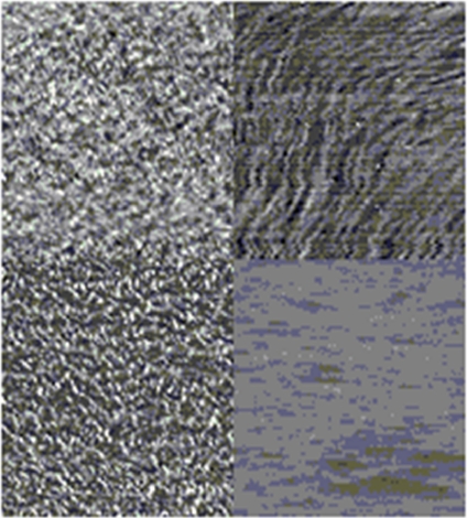
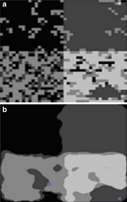
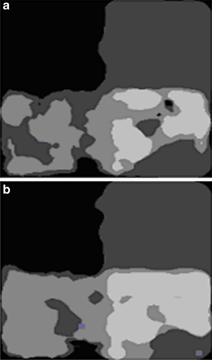
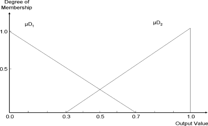
In this paper, a new neural-fuzzy approach is proposed for automated region segmentation in transrectal ultrasound images of the prostate. The goal of region segmentation is to identify suspicious regions in the prostate in order to provide decision support for the diagnosis of prostate cancer. The new automated region segmentation system uses expert knowledge as well as both textural and spatial features in the image to accomplish the segmentation. The textural information is extracted by two recurrent random pulsed neural networks trained by two sets of data (a suspicious tissues' data set and a normal tissues' data set). Spatial information is captured by the atlas-based reference approach and is represented as fuzzy membership functions. The textural and spatial features are synthesized by a fuzzy inference system, which provides a binary classification of the region to be evaluated.
Figures
Similar articles
-
A coarse-to-fine approach to prostate boundary segmentation in ultrasound images.Biomed Eng Online. 2005 Oct 11;4:58. doi: 10.1186/1475-925X-4-58. Biomed Eng Online. 2005. PMID: 16219098 Free PMC article. Clinical Trial.
-
Computer-aided prostate cancer detection using texture features and clinical features in ultrasound image.J Digit Imaging. 2008 Oct;21 Suppl 1(Suppl 1):S121-33. doi: 10.1007/s10278-008-9106-3. Epub 2008 Mar 6. J Digit Imaging. 2008. PMID: 18322751 Free PMC article.
-
Prostate tissue texture feature extraction for suspicious regions identification on TRUS images.J Digit Imaging. 2009 Oct;22(5):503-18. doi: 10.1007/s10278-008-9124-1. Epub 2008 May 13. J Digit Imaging. 2009. PMID: 18473140 Free PMC article.
-
A medical texture local binary pattern for TRUS prostate segmentation.Annu Int Conf IEEE Eng Med Biol Soc. 2007;2007:5605-8. doi: 10.1109/IEMBS.2007.4353617. Annu Int Conf IEEE Eng Med Biol Soc. 2007. PMID: 18003283
-
Temporal-based needle segmentation algorithm for transrectal ultrasound prostate biopsy procedures.Med Phys. 2010 Apr;37(4):1660-73. doi: 10.1118/1.3360440. Med Phys. 2010. PMID: 20443487
Cited by
-
Fuzzy logic: A "simple" solution for complexities in neurosciences?Surg Neurol Int. 2011 Feb 26;2:24. doi: 10.4103/2152-7806.77177. Surg Neurol Int. 2011. PMID: 21541006 Free PMC article.
References
-
- National Cancer Institute of Canada. Canadian cancer statistics 2008. Toronto, Canada, 2009
-
- Heidenreich A, Bolla M, Joniau S, van der Kwast TH, Matveev V, Mason, Mottet N, Schmid H-P, Wiegel T, Zattoni F. Guidelines on Prostate Cancer. Eur Urol 53(1):68–80, 2008 - PubMed
-
- Presti JC, Jr, O’Dowd GJ, Miller MC, Mattu R, Veltri RW. Extended peripheral zone biopsy schemes increase cancer detection rates and minimize variance in prostate specific antigen and age related cancer rates: results of a community multi-practice study. J Urol. 2003;169(1):125–129. doi: 10.1016/S0022-5347(05)64051-7. - DOI - PubMed
-
- Ethan J. Halpern, Stephen E. Strup. Using gray-scale and color and power doppler sonography to detect prostatic cancer. Am J Roentgenol 174:623–627, 2000 - PubMed
MeSH terms
LinkOut - more resources
Full Text Sources
Medical
