Review
. 2010 Aug;12(4):267-75.
doi: 10.1007/s11906-010-0118-2.

Endothelium-derived vasoactive factors and hypertension: possible roles in pathogenesis and as treatment targets

Affiliations
Review

Endothelium-derived vasoactive factors and hypertension: possible roles in pathogenesis and as treatment targets

Michel Félétou et al. Curr Hypertens Rep. 2010 Aug.

Abstract

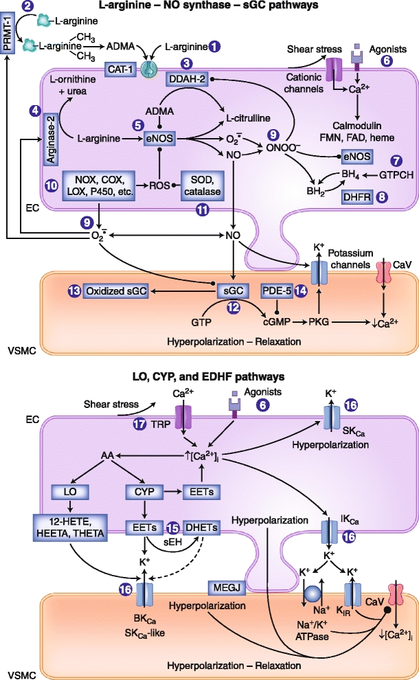
Endothelial cells regulate vascular tone by releasing various contracting and relaxing factors including nitric oxide (NO), arachidonic acid metabolites (derived from cyclooxygenases, lipoxygenases, and cytochrome P450 monooxygenases), reactive oxygen species, and vasoactive peptides. Additionally, another pathway associated with the hyperpolarization of the underlying smooth muscle cells plays a predominant role in resistance arteries. Endothelial dysfunction is a multifaceted disorder, which has been associated with hypertension of diverse etiologies, involving not only alterations of the L-arginine NO-synthase-soluble guanylyl cyclase pathway but also reduced endothelium-dependent hyperpolarizations and enhanced production of contracting factors, particularly vasoconstrictor prostanoids. This brief review highlights these different endothelial pathways as potential drug targets for novel treatments in hypertension and the associated endothelial dysfunction and end-organ damage.


Figures

Fig. 1
Fig. 1
Nitric oxide (NO) synthase, lipoxygenase (LO), and cytochrome P450 monooxygenase (CYP) pathways and responses mediated by endothelium-derived hyperpolarizing factors (EDHFs): potential sites of therapeutic intervention for hypertension. The circled numbers indicate potential sites of intervention: 1, L-arginine supplementation. 2, Inhibition of protein arginine N-methyltransferase type I (PRMT-I) to prevent the formation of asymmetric dimethyl-L-arginine (ADMA). 3, Increased expression and/or activity of dimethylarginine dimethylaminohydrolase-2 (DDAH-2) to facilitate ADMA catabolism. 4, Inhibition of arginase-2 to prevent L-arginine metabolism. 5, Increased expression and/or activity of endothelial nitric oxide synthase (eNOS). 6, Design of drugs that evoke endothelium-dependent relaxations. 7, Enhanced expression and/or activity of guanosine triphosphate cyclohydrolase (GTPCH), the rate-limiting enzyme for tetrahydrobiopterin (BH4) synthesis, or direct supplementation with BH4 or its precursor sepiapterin. 8, Enhanced expression and/or activity of dihydrofolate reductase (DHFR), involved in BH4 regeneration. 9, Scavengers of reactive oxygen species (ROS), antioxidants. 10, Inhibition of the activity and/or expression of enzymes that generate ROS, such as NAD(P)H oxidases (NOX), cyclooxygenases (COX), lipoxygenases (LOX), or cytochrome P450 monooxygenases (P450). 11, Enhanced expression and/or activity of enzymes that metabolize ROS, such as superoxide dismutase (SOD) or catalase (or, alternatively, synthesis of mimetics). 12, Stimulation of soluble guanylyl cyclase (sGC). 13, Activation of sGC. 14, Inhibition of phosphodiesterase-5 (PDE-5). 15, Inhibition of soluble epoxide hydrolase (sEH) to suppress degradation of epoxyeicosatrienoic acids (EETs). 16, Opening calcium-activated potassium channels of small, intermediate, or large conductance (SKCa, IKCa, BKCa). 17, Opening transient receptor potential channels (TRP). AA—arachidonic acid; BH2—dihydrobiopterin; CAT-1—cationic amino acid transporters; CaV—voltage-activated calcium channel; cGMP—cyclic guanosine monophosphate; DHETs—dihydroxyeicosatrienoic acids, EC—endothelial cell; FAD—flavin adenine dinucleotide; FMN—flavin mononucleotide; HEETA—hydroxy-epoxyeicosatrienoic acid; 12-HETE—12-hydroxyeicosatetraenoic acid; KIR—inward rectifying potassium channel; MEGJ—myoendothelial gap junction; O2•−—superoxide anion; ONOO—peroxynitrite; PKG—protein kinase G; THETA, trihydroxyeicosatrienoic acid; VSMC—vascular smooth muscle cell
Fig. 2
Fig. 2
Endothelium-dependent effects of acetylcholine in aorta of Wistar Kyoto (WKY) rats and spontaneously hypertensive rats (SHR). Left, Endothelium-dependent relaxations in normotensive WKY rats. Right, Cyclooxygenase-dependent, endothelium-dependent contractions to acetylcholine in SHR aorta. A23187—calcium ionophore; AA—arachidonic acid; AC—adenylyl cyclase; ATP—adenosine triphosphate; cAMP—cyclic adenosine monophosphate; CaV—voltage-activated calcium channel; cGMP—cyclic guanosine monophosphate; COX1—cyclooxygenase 1; eNOS—endothelial nitric oxide synthase; GTP—guanosine triphosphate; IP—PGI2 receptor; M—muscarinic receptor; O2•−—superoxide anion; PGH2—endoperoxide; PGI2—prostacyclin; PGIS—prostacyclin synthase; PLA2—phospholipase A2; P2Y—putinergic receptor Y2; sGC—soluble guanylyl cyclase; SR—sarcoplasmic reticulum; TP—TP receptor; TXA2—thromboxane A2. (From Félétou and Vanhoutte [17]. Used with permission of The American Physiological Society.)

References

    1. Furchgott RF, Zawadzki JV. The obligatory role of the endothelial cells in the relaxation of arterial smooth muscle by acetylcholine. Nature. 1980;288:373–376. doi: 10.1038/288373a0. - DOI - PubMed
    1. Vanhoutte PM. How we learned to say NO. Arteriosclerosis Thromb Vasc Biol. 2009;29:1156–1160. doi: 10.1161/ATVBAHA.109.190215. - DOI - PubMed
    1. Feletou M, Vanhoutte PM. EDHF: an update. Clin Sci (Lond) 2009;117:139–155. doi: 10.1042/CS20090096. - DOI - PubMed
    1. Vanhoutte PM, Tang EHC: Endothelium-dependent contractions: when a good guy turns bad! J Physiol 2008, 586:5295–5304. - PMC - PubMed
    1. Vanhoutte PM, Tang E, Félétou M, Shimokawa H. Endothelial dysfunction and vascular disease. Acta Physiol. 2009;196:193–222. doi: 10.1111/j.1748-1716.2009.01964.x. - DOI - PubMed