

Classical Article
doi: 10.1007/s11999-010-1416-3.
The classic: The treatment of chronic osteomyelitis with the maggot (larva of the blow fly). 1931
Affiliations
- PMID: 20532712
- PMCID: PMC3048276
- DOI: 10.1007/s11999-010-1416-3
Item in Clipboard
Classical Article
The classic: The treatment of chronic osteomyelitis with the maggot (larva of the blow fly). 1931
Clin Orthop Relat Res.
2011 Apr.
Abstract
This Classic article is a reprint of the original work by William S. Baer, MD, The Treatment of Chronic Osteomyelitis With the Maggot (Larva of the Blow Fly). An accompanying biographical sketch on William Baer, is available at DOI 10.1007/s11999-010-1415-4 . The Classic Article is ©1931 by the Journal of Bone and Joint Surgery, Inc. and is reprinted with permission from Baer WS. The treatment of chronic osteomyelitis with the maggot (larva of the blow fly). J Bone Joint Surg Am. 1931;13:438-475.
Figures

Showing male and female fly.

Full-grown maggots.

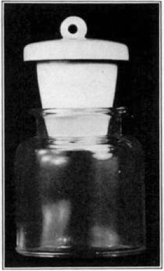
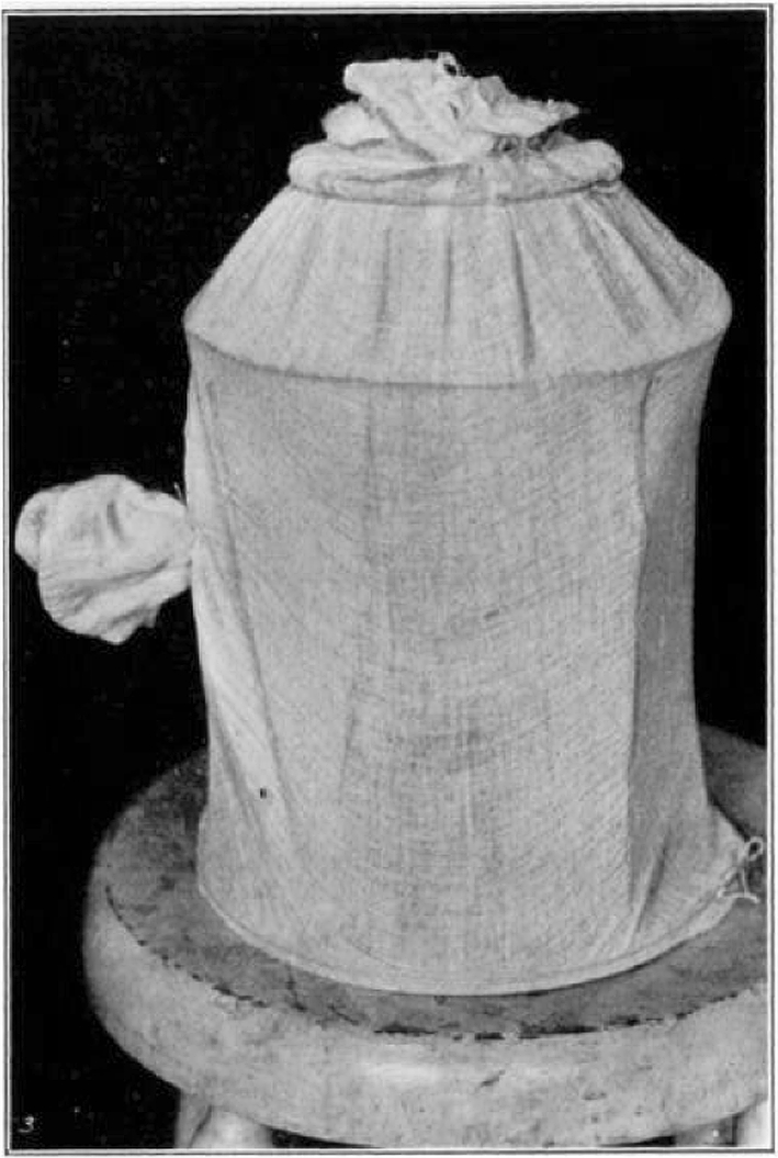
Container for sterile maggots.
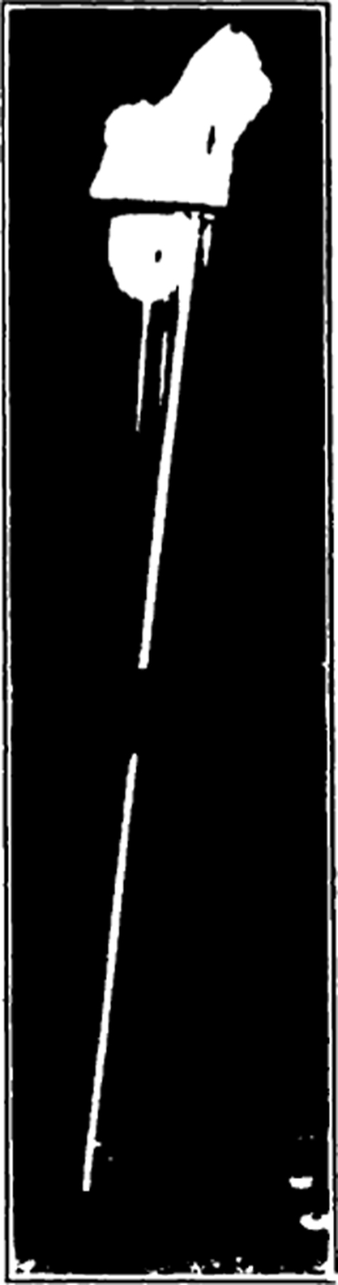
Test tube with applicator stick. Used as container when sterilizing the eggs.

Sterile food used in growing maggots for the patient’s wound.
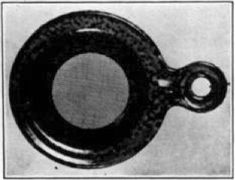
Gooch crucible. Complete for washing the eggs.
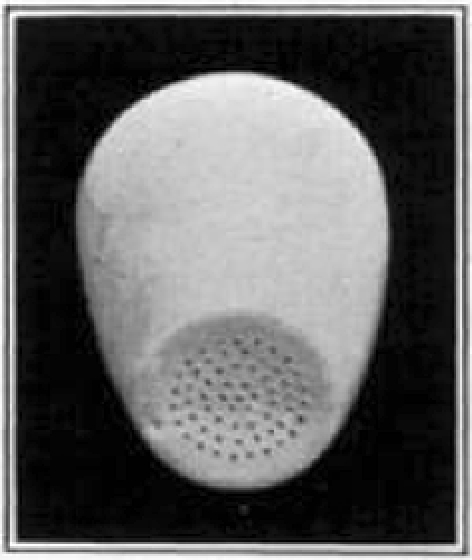
Gooch crucible showing perforations in the bottom.
Strainer used in washing maggots.
Cage for housing the flies. The side sleeve is used when feeding or catching the flies. When transferring the flies from one cage to another the top opening is untied and a clean cage is inverted over the old one. A light is placed over the top cage and the flies will at once fly into the clean cage. When they have all been captured the gauze can be drawn together and tied.
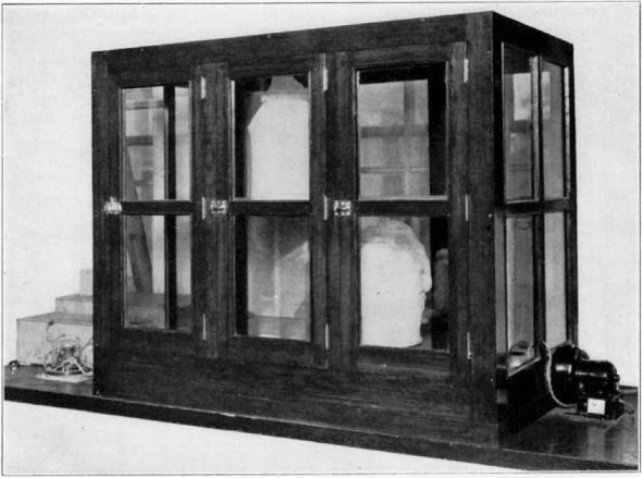
Incubator used for housing the flies. On the right the blower can be seen and on the left two boxes. The large box contains water to supply moisture and the smaller one contains the heating coils. On the left also is the thermostat control. The thermostat can be seen on the inside of the incubator on the right-hand side.

Showing cage turned back.

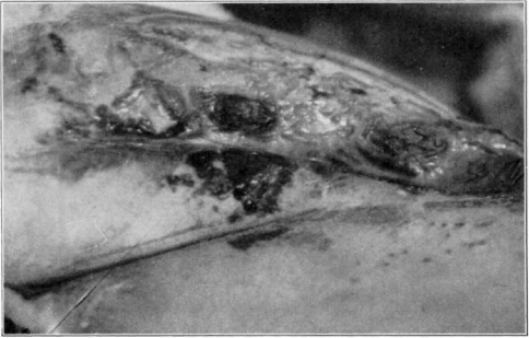
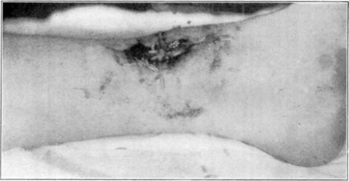
Showing maggots at work in wound.

Showing maggots at work in wound.

Showing maggots at work in wound.

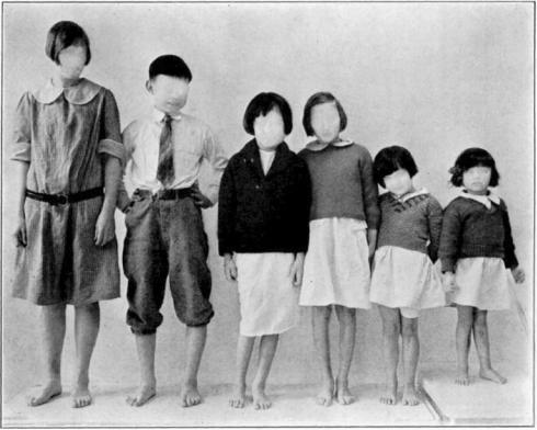
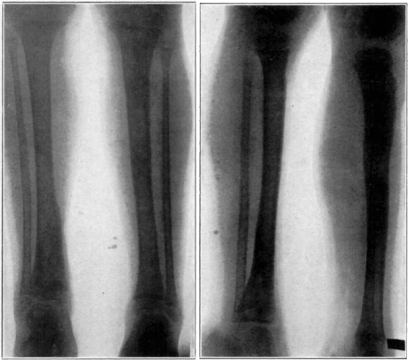
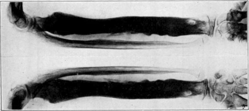
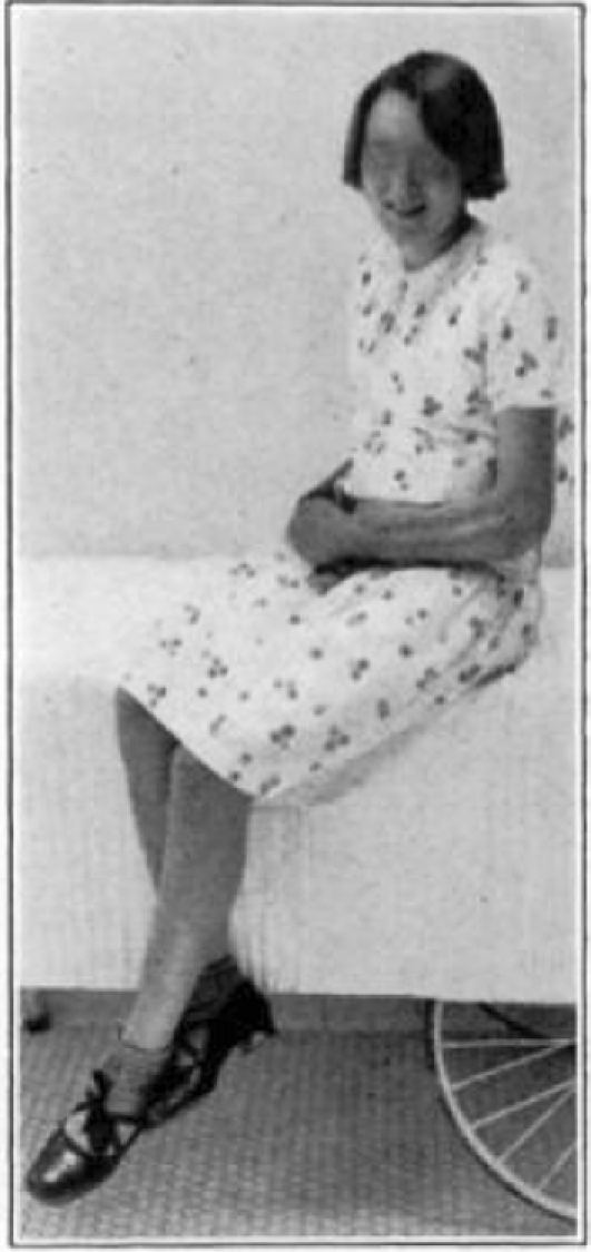
Cured cases of osteomyelitis following maggot treatment.
Another group of cases cured by the maggot treatment.
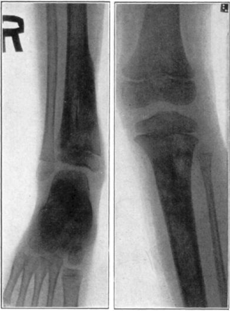
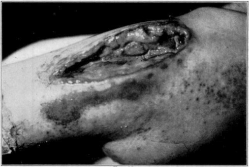
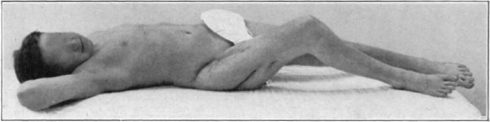
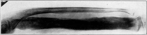
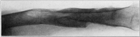
Case 1. B. M. Before operation.
Case 1. B. M. After operation.
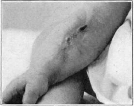
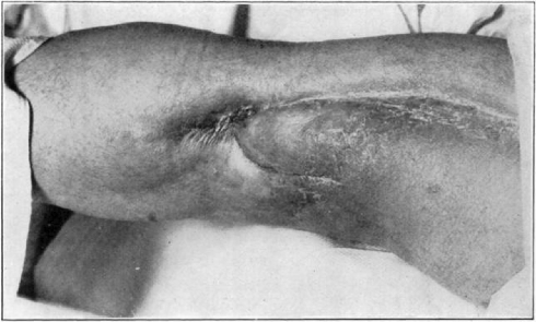
Case 1. B. M. Almost healed.
Case 1. B. M. Completely healed.

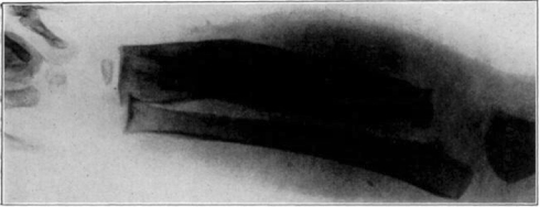
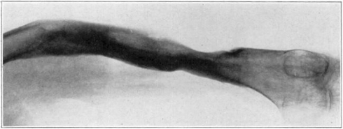
Case 3. A. W. Before operation.
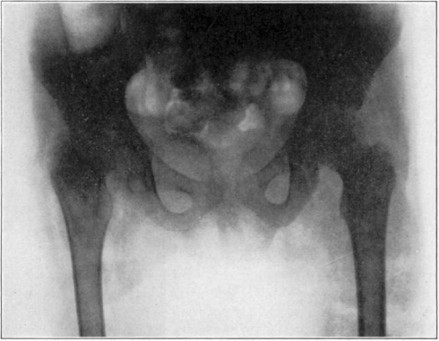
Case 3. A. W. After operation. Tuberculous hip.
Case 3. A. W. During treatment. Tuberculous hip.

Case 3. A. W. Almost healed. Tuberculous hip.

Case 3. A. W. After treatment. Tuberculous hip.

Case 3. A. W. Healed tuberculous hip.
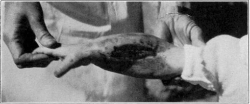
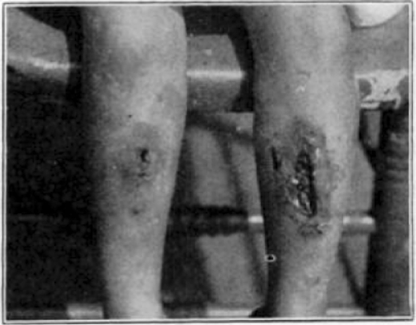
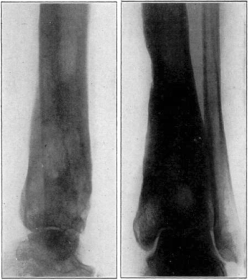
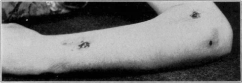
Case 4. B. C. Before operation.

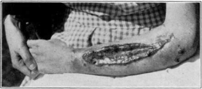
Case 4. B. C. After operation.
Case 4. B. C. During treatment.
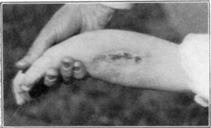
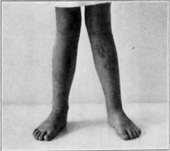
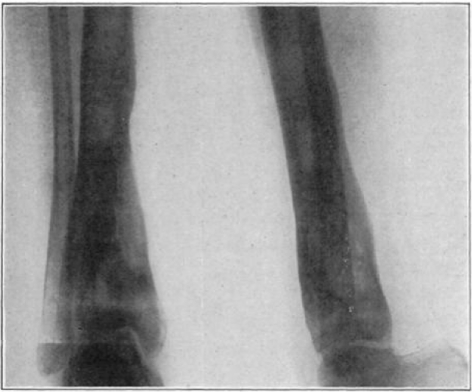
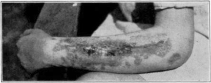
Case 4. B. C. Almost healed.
Case 4. B. C. Healed.

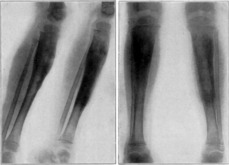
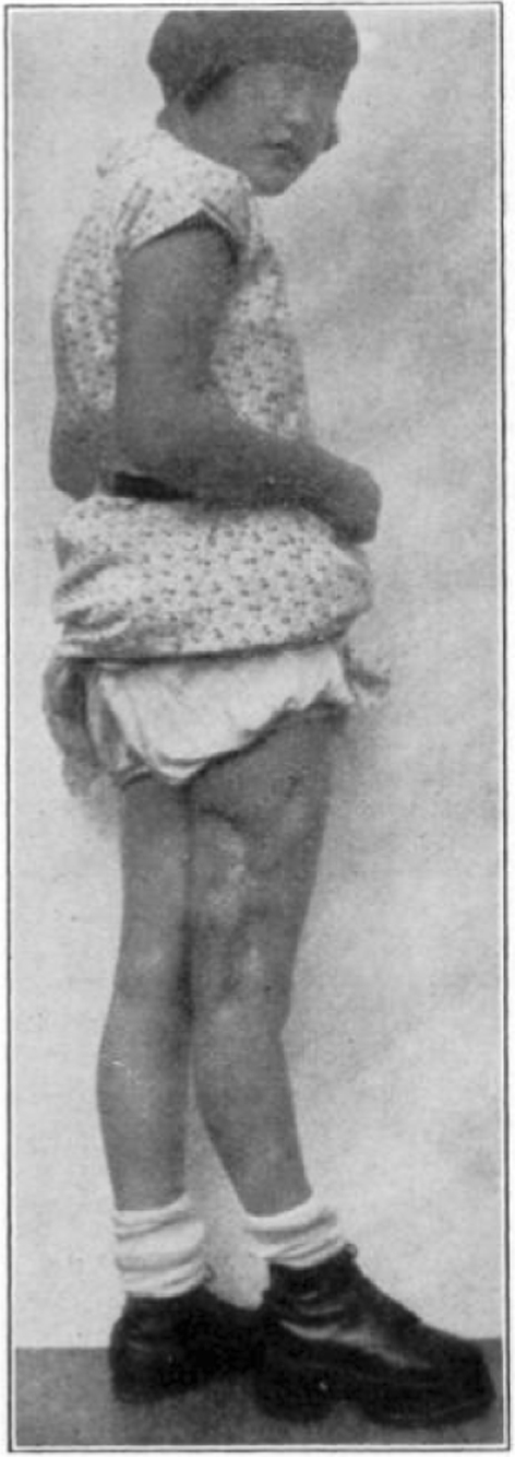
Case 15. F. R. Before operation.
Case 15. F. R. During treatment.

Case 15. F. R. During treatment.

Case 15. F. R. Almost healed.
Case 15. F. R. Healed osteomyelitis.

Case 20. L. P. Before operation.
Case 20. L. P. After operation.
Case 20. L. P. During treatment.
Case 20. L. P. Completely healed.
Case 23. E. W. Before operation.
Case 23. E. W. After operation.

Case 23. E. W. Completely healed.
Case 23. E. W. Showing method of treatment.
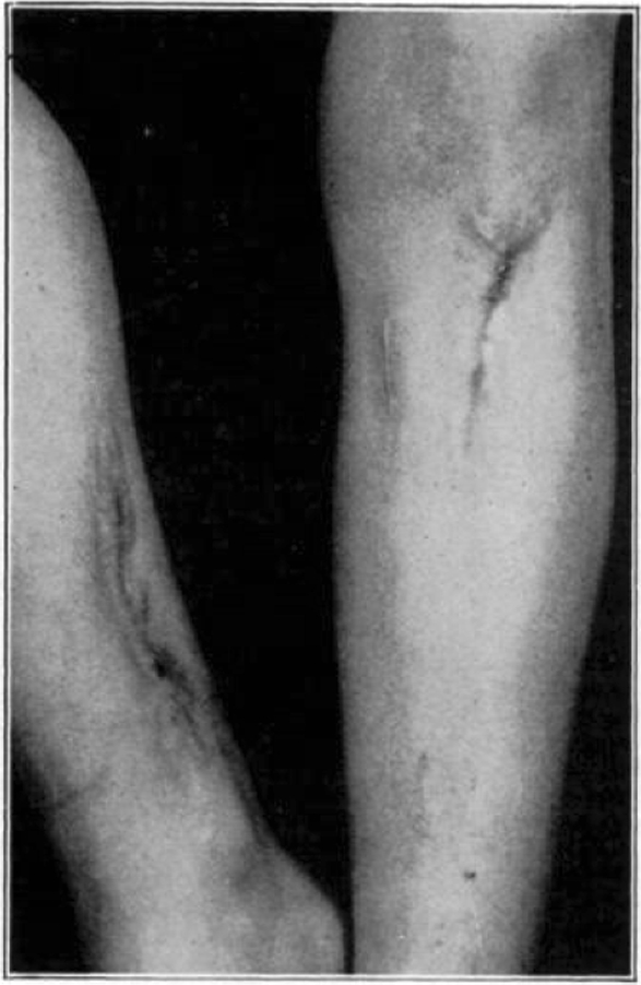
Case 25. R. Y. Healed osteomyelitis.
Case 25. R. Y. Healed osteomyelitis.
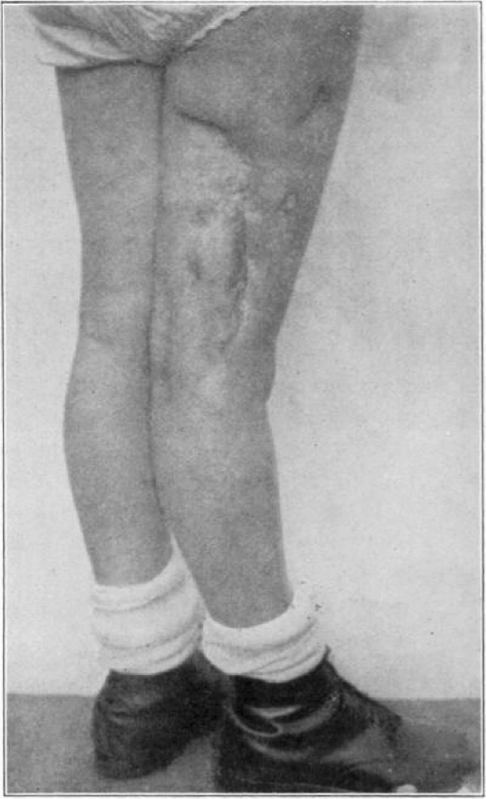
Case 28. R. L. Before operation.
Case 28. R. L. After operation.
Case 28. R. L. Before treatment.
Case 28. R. L. During treatment.
Case 28. R. L. Nearly healed.
Case 28. R. L. Healed.
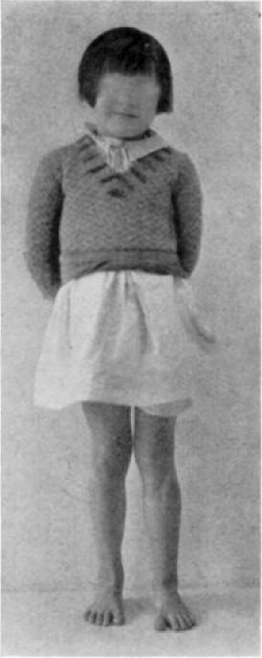
Case 57. B. G. Nine years’ duration. Seven previous operations. Seven operations. Healed.

Case 57. B. G. Three months after operation.
Case 57. B. G. Three months after operation.
Case 57. B. G. Ten months after operation. Nine years’ duration. Seven previous operations. Seven operations. Healed.
Publication types
MeSH terms
Personal name as subject
- Actions
LinkOut - more resources
Full Text Sources
Other Literature Sources
