Primary diffuse large B-cell lymphoma of the oral cavity: germinal center classification
- PMID: 20533006
- PMCID: PMC2923304
- DOI: 10.1007/s12105-010-0184-4
Primary diffuse large B-cell lymphoma of the oral cavity: germinal center classification
Abstract
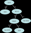
Primary lymphomas of the oral cavity are rare and the most frequent type is diffuse large B-cell lymphoma (DLBCL). Recently, several reports have highlighted the value of classifying DLBCL into prognostically important subgroups, namely germinal center B-cell like (GCB) and non-germinal center B-cell like (non-GCB) lymphomas based on gene expression profiles and by immunohistochemical expression of CD10, BCL6 and MUM-1. GCB lymphomas tend to exhibit a better prognosis than non-GCB lymphomas. Studies validating this classification have been done for DLBCL of the breast, CNS, testes and GI tract. Therefore we undertook this study to examine if primary oral DLBCLs reflect this trend. We identified 13 cases (age range 38-91 years) from our archives dating from 2003-09. IHC was performed using antibodies against germinal center markers (CD10, BCL6), activated B-cell markers (MUM1, BCL2) and Ki-67 (proliferation marker). Cases were sub-classified as GCB subgroup if CD10 and/or BCL6 were positive and MUM-1, was negative and as non-GCB subgroup if CD10 was negative and MUM-1 was positive. Immunoreactivity was noted in 2/13 cases for CD10, in 12/13 for BCL6, in 8/13 for MUM-1, and in 6/13 for BCL2. Therefore, 8/13 (58%) were sub-classified as non-GCB DLBCLs and 5/13 (42%) as GCB subgroup. All tumors showed frequent labeling with Ki-67 (range 40-95%). Four of the 8 patients with non-GCB subgroup succumbed to their disease, with the mean survival rate of 16 months. Two patients in this group are alive, one with no evidence of disease and another with disease. No information was available for the other 3 patients in this group. Four of the 5 patients in the GCB subgroup were alive with no evidence of disease and one patient succumbed to complications of therapy and recurrent disease after 18 months. In conclusion, our analysis shows that primary oral DLBCL predominantly belongs to the non-GCB subgroup, which tends to exhibit a poorer prognosis. These findings could allow pathologists to provide a more accurate insight into the potential aggressive behavior and poorer prognosis of these lymphomas.
Figures


References
-
- Swerdlow SH, Campo E, Harris NL, Jaffe ES, Pileri SA, Stein H, et al. WHO Classification of Tumours of Haematopoietic and Lymphoid Tissues. 4. Lyon: IARC Press; 2008.
-
- Kolokotronis A, Konstantinou N, Christakis I, Papadimitriou P, Matiakis A, Zaraboukas T, et al. Localized B-cell non-Hodgkin’s lymphoma of oral cavity and maxillofacial region: a clinical study. Oral Surg Oral Med Oral Pathol Oral Radiol Endod. 2005;99(3):303–310. doi: 10.1016/j.tripleo.2004.03.028. - DOI - PubMed
Publication types
MeSH terms
Substances
LinkOut - more resources
Full Text Sources
Medical

