. 2010 Aug 1;518(15):3046-64.
doi: 10.1002/cne.22379.

Age changes in myelinated nerve fibers of the cingulate bundle and corpus callosum in the rhesus monkey

Affiliations

Age changes in myelinated nerve fibers of the cingulate bundle and corpus callosum in the rhesus monkey

Michael P Bowley et al. J Comp Neurol. .

Abstract

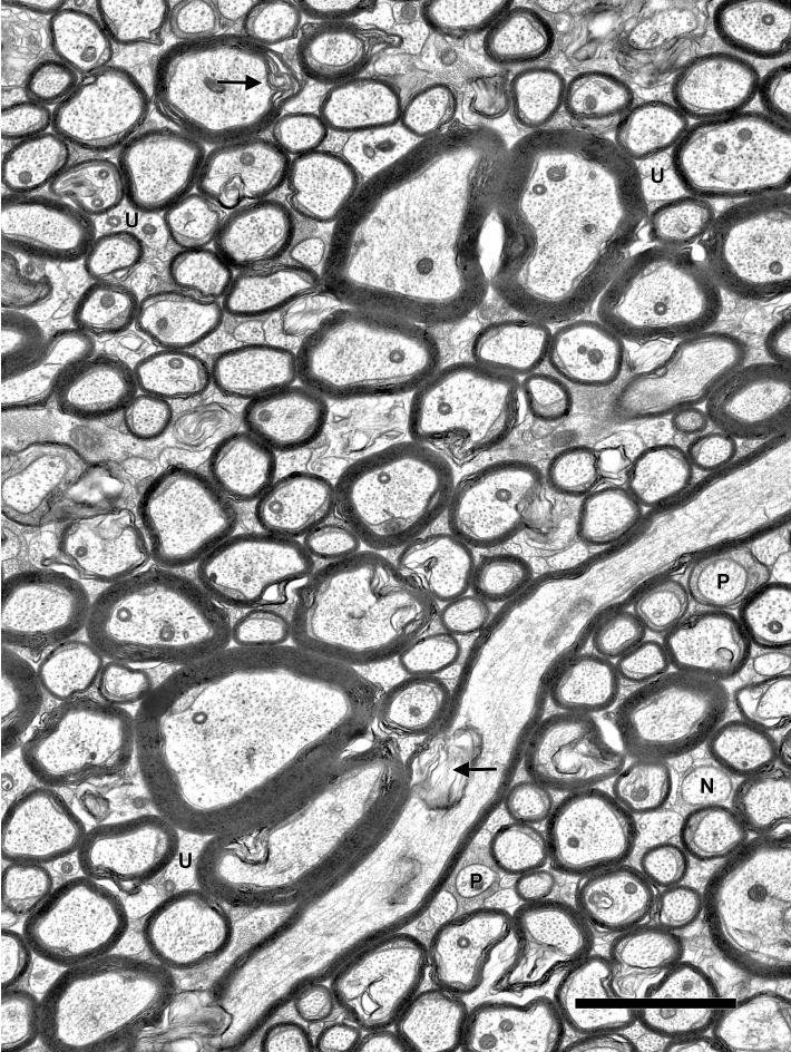
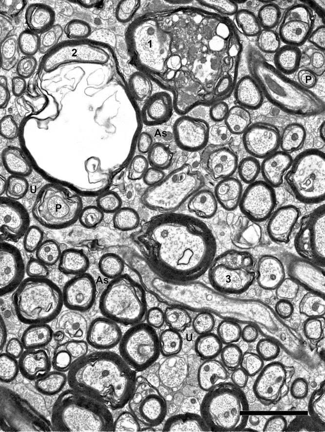
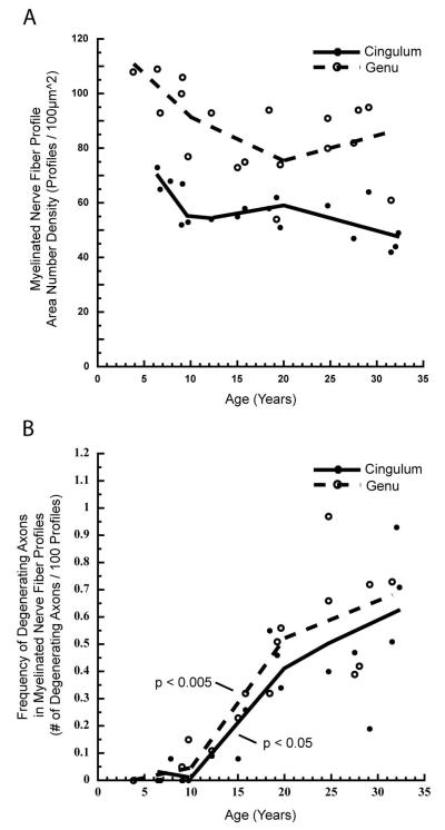
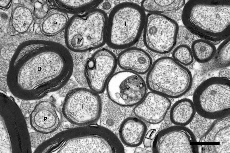
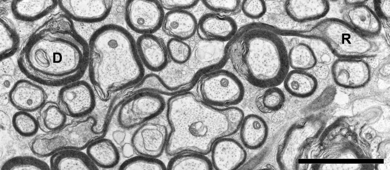
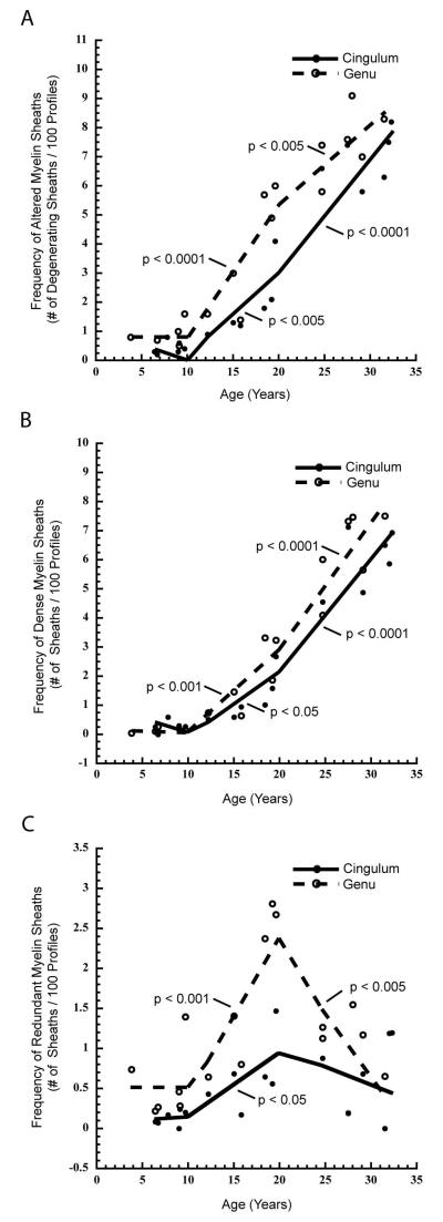
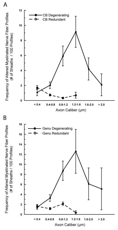
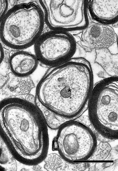
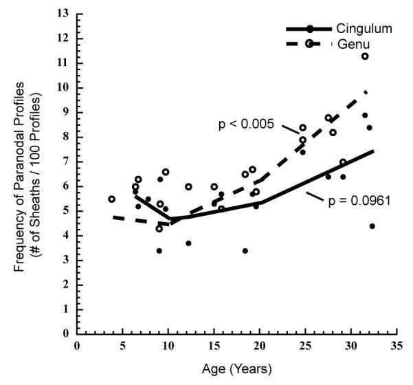
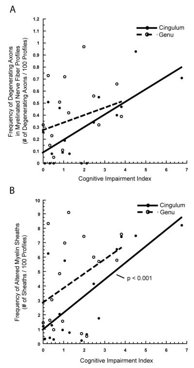
Aging is accompanied by deficits in cognitive function, which may be related to the vulnerability of myelinated nerve fibers to the normal process of aging. Loss of nerve fibers, together with age-related alterations in myelin sheath structure, may result in the inefficient and poorly coordinated conduction of neuronal signals. Until now, the ultrastructural analysis of cerebral white matter fiber tracts associated with frontal lobe areas critical in cognitive processing has been limited. In this study we analyzed the morphology and area number density of myelinated nerve fibers in the cingulate bundle and genu of the corpus callosum in behaviorally assessed young, middle aged, and old rhesus monkeys (Macaca mulatta). In both structures, normal aging results in a 20% decrease in the number of myelinated nerve fibers per unit area, while remaining nerve fibers exhibit an increasing frequency of degenerative changes in their myelin sheaths throughout middle and old age. Concomitantly, myelination continues in older monkeys, suggesting ongoing, albeit inadequate, reparative processes. Despite similar patterns of degeneration in both fiber tracts, only the age-related changes in the cingulate bundle correlate with declining cognitive function, underscoring its role as a critical corticocortical pathway linking the medial prefrontal, cingulate, and parahippocampal cortices in processes of working memory, recognition memory, and other higher cognitive faculties. These results further demonstrate the important role myelinated nerve fiber degeneration plays in the pathogenesis of age-related cognitive decline.


Figures

Figure 1
Figure 1
This figure depicts the cross-sectional areas for the cingulate white matter (A and B) in the coronal plane, and the genu of the corpus callosum (C and D) in the mid-sagittal plane. A: The cingulate gyrus and underlying white matter at the level of the anterior commissure. The cingulate white matter (cwm) is bounded dorsally, medially, and laterally by the cingulate cortex (cg). Ventrally, it is bounded by the body of the corpus callosum (cc). An inferolateral boundary was arbitrarily defined by a line oriented perpendicular to the cingulate sulcus (cs), from the gray matter/white matter boundary dorsally, to the underlying corpus callosum (cc) ventrally. B: Enlarged diagram of the cingulate white matter from A. The cingulate white matter is composed of both the cingulate bundle proper (cb) and local system fibers (u) underlying the cingulate cortex (cg). The hatched area represents the portion of the cingulate bundle sampled for electron microscopic analysis. C: A mid-sagittal diagram of the corpus callosum of the rhesus monkey. The genu (g) is the anterior-most portion of the corpus callosum. Its posterior boundary is defined by a straight line, perpendicular to the long axis of the corpus callosum, at the boundary of the corpus callosum with the anterior tip of the septum pellucidum (sep). Splenium (s), body of the corpus callosum (b), rostrum (r). D: An enlarged diagram of the mid-sagittal area of genu of the corpus callosum in C. The hatched area represents the dorsal portion of the genu sampled for electron microscopic analysis.
Figure 2
Figure 2
Electron micrograph of the cingulate bundle of a 9-year-old female rhesus monkey (AM096), showing the tightly packed myelinated nerve fibers. The myelin sheaths of some nerve fibers show shearing defects (arrows), an artifact of tissue processing. Interspersed amongst these myelinated nerve fibers, either singly or in clusters, are unmyelinated nerve fibers (U), as well as nodal (N) and paranodal profiles (P) of myelinated nerve fibers. Scale bar = 2μm.
Figure 3
Figure 3
Electron micrograph of the cingulate bundle of a 24-year-old female rhesus monkey (AM100). The myelinated nerve fibers are less tightly packed than in younger monkeys (Fig. 2), and astrocytic processes (As) are more frequent between adjacent nerve fibers. Some nerve fibers (1 and 3) contain dense cytoplasm in splits of their myelin sheaths. Another myelinated nerve fiber (2) shows ballooning of the sheath. Interspersed amongst these myelinated nerve fibers are unmyelinated nerve fibers (U). Profiles through paranodes (P) are also indicated. Scale bar = 2μm.
Figure 4
Figure 4
Scatterplots with piecewise, linear fits showing: A) The number of myelinated nerve fibers per unit area, and B) the frequency of myelinated nerve fiber profiles with degenerating axons in the cingulate bundle and genu of the corpus callosum of young (<10 years), middle aged (10-20 years), and old (>20 years) rhesus monkeys. In both structures, a decrease in myelinated nerve fibers occurs gradually with age, with none of the three age groups exhibiting a significant change in area number density (A). The frequency of degenerating myelinated axons significantly increases only during middle age in both the genu (p < 0.005) and cingulate bundle (p < 0.05; B).
Figure 5
Figure 5
Electron micrograph from the cingulate bundle of a 31 year-old male rhesus monkey (AM091). Internodal (I) and paranodal (P) profiles of myelinated nerve fibers are indicated. The axon of one myelinated nerve fiber (asterisk) is degenerating. Another myelinated nerve fiber (D) is surrounded by sheath containing dense cytoplasm. Scale bar= 1μm.
Figure 6
Figure 6
Electron micrograph of the cingulate bundle of an 18.4 year-old female rhesus monkey (AM 221). One axon (R), is ensheathed by a myelin sheath too large for the axon, referred to as a redundant sheath, while a second axon (D) is enclosed by a sheath with a split at the major dense line, referred to as a dense sheath Scale bar = 2μm.
Figure 7
Figure 7
Scatterplots with a piecewise, linear fits showing: A) the frequency of myelinated nerve fiber profiles with altered myelin sheaths (including dense sheaths, redundant sheaths, and myelin balloons), B) the frequency of myelinated nerve fiber profiles with dense myelin sheaths, and C) the frequency of myelinated nerve fiber profiles with redundant myelin sheaths in the cingulate bundle and the genu of the corpus callosum of young (<10 years), middle aged (10-20 years), and old (>20 years) rhesus monkeys. As shown in A, a significant increase in the frequency of total altered myelin sheaths is seen in middle and old age groups in both the genu (middle age, p < 0.0001; old, p < 0.005) and cingulate bundle (middle age, p < 0.005; old, p < 0.0001). Likewsie, in B, a significant increase in the frequency of dense myelin sheaths is seen in middle and old age groups in both the genu (middle age, p < 0.001; old, p < 0.0001) and cingulate bundle (middle age, p < 0.05; old, p < 0.0001). In C, a significant increase in the frequency of redundant myelin sheaths is evident in middle age monkeys in both the genu (p < 0.001) and cingulate bundle (p < 0.05). Additionally, there is a significant decrease in the frequency of redundant myelin sheaths in the genu of old aged monkeys (p < 0.005) but not in the cingulate bundle.
Figure 8
Figure 8
Line graphs showing the frequency of degenerating (dense sheaths and balloons) and of redundant myelin sheaths in the cingulate bundle (A) and genu of the corpus callosum (B) as a function of mean axon diameter in monkeys over 10 years of age. Nerve fibers are divided into 6 size classes from < 0.4μm to > 2.0μm in diameter.
Figure 9
Figure 9
Electron micrograph of the cingulate bundle of a 31-year-old male rhesus monkey (AM091). The indicated axon (asterisk) is thinly myelinated. Scale bar = 1μm
Figure 10
Figure 10
Scatterplot with a piecewise, linear fit showing the frequency of myelinated nerve fiber profiles through paranodes in the cingulate bundle and genu of the corpus callosum of young (<10 years), middle aged (10-20 years), and old (>20 years) monkeys. A change in the frequency of paranodal profiles is most prominent in old age, significantly increasing in the genu of the corpus callosum (p < 0.005), and exhibiting a trend toward a significant increase in the cingulate bundle (p = 0.0961) of old monkeys. No significant age-group specific changes in paranodal frequency are evident in either structure in young or middle aged monkeys.
Figure 11
Figure 11
A) Scatterplot with a linear fit, showing the frequency of myelinated nerve fiber profiles with degenerating axons in the cingulate bundle and genu of the corpus callosum versus a measure of global cognitive impairment, the cognitive impairment index (CII). An increase in CII, indicating worsening cognitive function, is associated with an increase in the frequency of degenerating axons in the cingulate bundle (p < 0.005) but not in the genu of the corpus callosum (p = 0.2465). B) Scatterplot with a linear fit, showing the frequency of myelinated nerve fiber profiles with altered myelin sheaths in the cingulate bundle and genu of the corpus callosum versus CII. Worsening cognitive function is significantly associated with an increased frequency of altered myelin sheaths in the cingulate bundle (p < 0.001) but not the genu (p = 0.1086).

References

    1. Albert MS. Cognitive Function. In: Albert MS, Moss MB, editors. Geriatric Neuropsychology. The Guilford Press; New York: 1988. pp. 33–53.
    1. Albert MS. Neuropsychological and neurophysiological changes in healthy adult humans across the age range. Neurobiol Aging. 1993;14:623–625. - PubMed
    1. Allen JS, Bruss J, Brown CK, Damasio H. Normal neuroanatomical variation due to age: the major lobes and a parcellation of the temporal region. Neurobiol Aging. 2005;26:1245–1260. discussion 1279-1282. - PubMed
    1. Barbas H, Pandya DN. Topography of commissural fibers of the prefrontal cortex in the rhesus monkey. Exp Brain Res. 1984;55:187–191. - PubMed
    1. Blakemore WF. Observations on oligodendrocyte degeneration, the resolution of status spongiosus and remyelination in cuprizone intoxication in mice. J Neurocytol. 1972;1:413–426. - PubMed

Publication types

MeSH terms