Review
. 2010 Aug 1;88(10):2083-90.
doi: 10.1002/jnr.22392.

Synapse loss in dementias

Affiliations
Review

Synapse loss in dementias

Ryan Clare et al. J Neurosci Res. .

Abstract

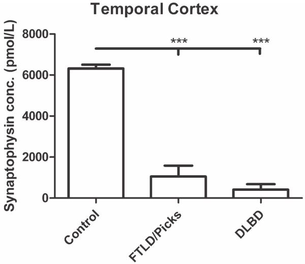
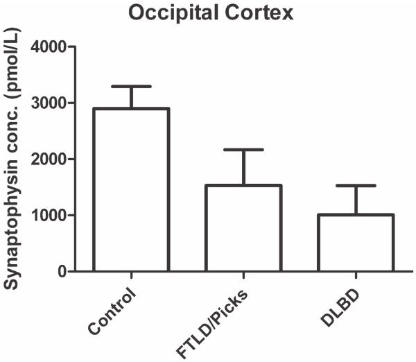
Synaptic transmission is essential for nervous system function, and its dysfunction is a known major contributing factor to Alzheimer's-type dementia. Antigen-specific immunochemical methods are able to characterize synapse loss in dementia through the quantification of various synaptic proteins involved in the synaptic cycle. These immunochemical methods applied to the study of Alzheimer's disease (AD) brain specimens have correlated synaptic loss with particularly toxic forms of amyloid-beta protein and have also established synapse loss as the best correlate of dementia severity. A significant but comparatively circumscribed amount of literature describes synaptic decline in other forms of dementia. Ischemic vascular dementia (IVD) is quite heterogeneous, and synapse loss in IVD seems to be variable among IVD subtypes, probably reflecting its variable neuropathologic correlates. Loss of synaptic protein has been identified in vascular dementia of the Binswanger type and Spatz-Lindenberg's disease. Here we demonstrate a significant loss of synaptophysin density within the temporal lobe of frontotemporal dementia (FTD) patients.


Figures

Fig. 1
Fig. 1
Synaptophysin protein distribution in tissue sections of neocortex and subcortical white matter from frontal (A,B,D,E) and superior temporal (C,F) lobes from patients with three different diseases: Alzheimer’s disease (AD; A,D), Pick’s disease (PD; B,E), and AD with severe cerebral amyloid angiopathy (CAA; C,F) illustrated with immunohistochemistry displayed at low (A–C) and high (D–F) magnification. Scale bars = 1 mm in C (applies to A–C); 0.5 mm in F (applies to D–F).
Fig. 2
Fig. 2
Sandwich ELISA results for temporal lobe samples from FTD, control, and DLBD patients. Autopsy tissue was acquired from the Alzheimer’s Disease Research Center (ADRC) Brain Bank at the University of California, Los Angeles. Three FTLD brains, one Pick’s disease brain, and five DLBD brains were selected, together with four age-matched control brains. Upon thawing, 0.4 g of temporal lobe gray matter was dissected and homogenized on ice. Lysates were centrifuged and stored at −20°C. The 96-well plate was coated with monoclonal mouse synaptophysin (Chemicon, Temecula, CA) diluted 1:1,000. After the wells had been blocked with 5% nonfat dry milk-0.2% Tween-20 blocking buffer, the lysates were added. The polyclonal rabbit second primary antibody (Dako, Glostrup, Denmark) diluted 1:400 was then added, followed by the goat anti-rabbit alkaline phosphatase-conjugated secondary antibody (Invitrogen, Carlsbad, CA) at a 1:1,000 dilution. Measurements were taken at 415 nm using a Bio-Rad Benchmark microplate reader 10 min after the Alkaline Phosphatase Substrate Kit (Bio-Rad, Hercules, CA) had been added. There was no correlation between PMI and synaptophysin concentration (correlation coefficient of 0.394). ***P < 0.001 (one-way ANOVA, Bonferroni’s multiple-comparisons test).
Fig. 3
Fig. 3
Sandwich ELISA results for occipital lobe samples from FTD, control, and DLBD patients. Three FTLD brains, two Pick’s disease brains, and five DLBD brains were selected, together with four age-matched control brains. Occipital lobe cortex samples were processed according to the protocol described for temporal lobe cortex in the legend to Figure 2. No statistically significant difference was found between the groups, and there was no correlation between PMI and synaptophysin concentration (correlation coefficient of 0.0061).

Similar articles

Cited by

References

    1. Bergeron C, Ranalli PJ, Miceli PN. Amyloid angiopathy in Alzheimer’s disease. Can J Neurol Sci. 1987;14:564–569. - PubMed
    1. Blennow K, Bogdanovic N, Alafuzoff I, Ekman R, Davidsson P. Synaptic pathology in Alzheimer’s disease: relation to severity of dementia, but not to senile plaques, neurofibrillary tangles, or the ApoE4 allele. J Neural Transm. 1996;103:603–618. - PubMed
    1. Brown DF, Risser RC, Bigio EH, Tripp P, Stiegler A, Welch E, Eagan KP, Hladik CL, White CL., III Neocortical synapse density and Braak stage in the lewy body variant of Alzheimer disease: a comparison with classic Alzheimer disease and normal aging. J Neuropathol Exp Neurol. 1998;57:955–960. - PubMed
    1. Brun A, Liu X, Erikson C. Synapse loss and gliosis in the molecular layer of the cerebral cortex in Alzheimer’s disease and in frontal lobe degeneration. Neurodegeneration. 1995;4:171–177. - PubMed
    1. Buttini M, Masliah E, Barbour R, Grajeda H, Motter R, Johnson-Wood K, Khan K, Seubert P, Freedman S, Schenk D, Games D. Beta-amyloid immunotherapy prevents synaptic degeneration in a mouse model of Alzheimer’s disease. J Neurosci. 2005;25:9096–9101. - PMC - PubMed

Publication types