

Dissociable brain states linked to common and creative object use
- PMID: 20533561
- PMCID: PMC3846690
- DOI: 10.1002/hbm.21056
Dissociable brain states linked to common and creative object use
Abstract
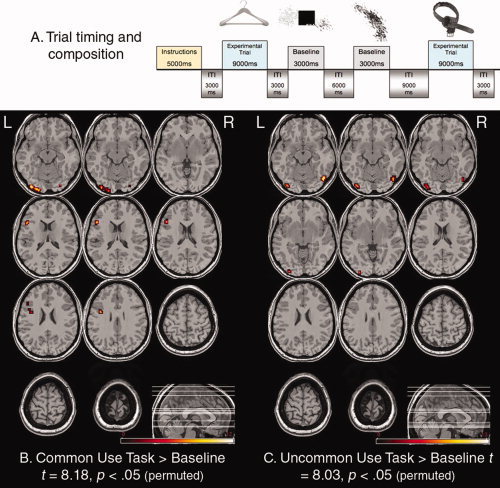
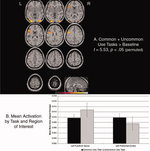
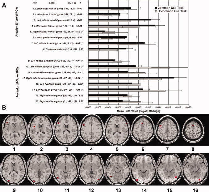
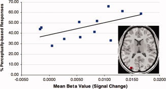
Studies of conceptual processing have revealed that the prefrontal cortex is implicated in close-ended, deliberate memory retrieval, especially the left ventrolateral prefrontal regions. However, much of human thought-particularly that which is characterized as creative-requires more open-ended, spontaneous memory retrieval. To explore the neural systems that support conceptual processing under these two distinct circumstances, we obtained functional magnetic resonance images from 24 participants either while retrieving the common use of an everyday object (e.g., "blowing your nose," in response to a picture of a tissue) or while generating a creative (i.e., uncommon but plausible) use for it (e.g., "protective padding in a package"). The patterns of activation during open- and closed-ended tasks were reliably different, with regard to the magnitude of anterior versus posterior activation. Specifically, the close-ended task (i.e., Common Use task) reliably activated regions of lateral prefrontal cortex, whereas the open-ended task (i.e., Uncommon Use task) reliably activated regions of occipito-temporal cortex. Furthermore, there was variability across subjects in the types of responses produced on the open-ended task that was associated with the magnitude of activation in the middle occipital gyrus on this task. The present experiment is the first to demonstrate a dynamic tradeoff between anterior frontal and posterior occipitotemporal regions brought about by the close- or open-ended task demands.
Copyright © 2010 Wiley-Liss, Inc.
Figures
References
-
- Aguire GK, Zarahn E, D'Esposito M ( 1998): The variability of human BOLD hemodynamic responses. Neuroimage 8: 360–369. - PubMed
-
- Alexander JK, Hillier A, Smith RM, Tivarus ME, Beversdorf DO ( 2007): Beta‐adrenergic modulation of cognitive flexibility during stress. J Cogn Neurosci 19: 468–478. - PubMed
-
- Apperly IA, Samson D, Chiavarino C, Humphreys GW ( 2004): Frontal and temporo‐parietal lobe contributions to theory of mind: Neuropsychological evidence from a false belief task with reduced language and executive demands. J Cogn Neurosci 16: 1773–1784. - PubMed
-
- Badre D, Wagner AD ( 2007): Left ventrolateral prefrontal cortex and the cognitive control of memory. Neuropsychologia 45: 2883–1901. - PubMed
-
- Braver TS, Hannes R ( 2006): Functional neuroimaging of executive functions In: Cabeza R, Kingston A, editors. Handbook of Functional Neuroimaging of Cognition, 2nd ed. Cambridge, MA: MIT Press; pp 307–348.
Publication types
MeSH terms
Grants and funding
LinkOut - more resources
Full Text Sources
