

Microstructural development: organizational differences of the fiber architecture between children and adults in dorsal and ventral visual streams
- PMID: 20533564
- PMCID: PMC6869943
- DOI: 10.1002/hbm.21080
Microstructural development: organizational differences of the fiber architecture between children and adults in dorsal and ventral visual streams
Abstract
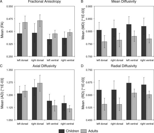
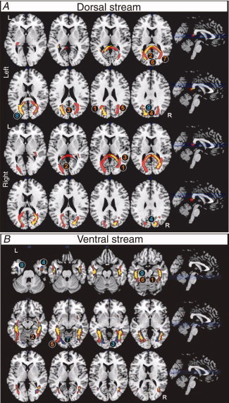
Visual perceptual skills are basically mature by the age of 7 years. White matter, however, continues to develop until late adolescence. Here, we examined children (aged 5-7 years) and adults (aged 20-30 years) using diffusion tensor imaging (DTI) fiber tracking to investigate the microstructural maturation of the visual system. We characterized the brain volumes, DTI indices, and architecture of visual fiber tracts passing through white matter structures adjacent to occipital and parietal cortex (dorsal stream), and to occipital and temporal cortex (ventral stream). Dorsal, but not ventral visual stream pathways were found to increase in volume during maturation. DTI indices revealed expected maturational differences, manifested as decreased mean and radial diffusivities and increased fractional anisotropy in both streams. Additionally, fractional anisotropy was increased and radial diffusivity was decreased in the adult dorsal stream, which can be explained by specific dorsal stream myelination or increasing fiber compaction. Adult dorsal stream architecture showed additional intra- and interhemispheric connections: Dorsal fibers penetrated into contralateral hemispheres via commissural structures and projection fibers extended to the superior temporal gyrus and ventral association pathways. Moreover, intra-hemispheric connectivity was particularly strong in adult dorsal stream of the right hemisphere. Ventral stream architecture also differed between adults and children. Adults revealed additional connections to posterior lateral areas (occipital-temporal gyrus), whereas children showed connections to posterior medial areas (posterior parahippocampal and lingual gyrus). Hence, in addition to dorsal stream myelination or fiber compaction, progressing maturation of intra- and interhemispheric connectivity may contribute to the development of the visual system.
Copyright © 2010 Wiley-Liss, Inc.
Figures
Similar articles
-
Neurodevelopment of the visual system in typically developing children.Prog Brain Res. 2011;189:113-36. doi: 10.1016/B978-0-444-53884-0.00021-X. Prog Brain Res. 2011. PMID: 21489386 Review.
-
Blindness alters the microstructure of the ventral but not the dorsal visual stream.Brain Struct Funct. 2016 Jul;221(6):2891-903. doi: 10.1007/s00429-015-1078-8. Epub 2015 Jul 2. Brain Struct Funct. 2016. PMID: 26134685
-
The Relationship between White Matter and Reading Acquisition, Refinement and Maintenance.Dev Neurosci. 2018;40(3):209-222. doi: 10.1159/000489491. Epub 2018 Jun 25. Dev Neurosci. 2018. PMID: 29940596
-
Fiber tracts of the dorsal language stream in the human brain.J Neurosurg. 2016 May;124(5):1396-405. doi: 10.3171/2015.5.JNS15455. Epub 2015 Nov 20. J Neurosurg. 2016. PMID: 26587654
-
Fiber anatomy of dorsal and ventral language streams.Brain Lang. 2013 Nov;127(2):192-204. doi: 10.1016/j.bandl.2012.04.015. Epub 2012 May 24. Brain Lang. 2013. PMID: 22632814 Review.
Cited by
-
Effect of number of diffusion-encoding directions in diffusion metrics of 5-year-olds using tract-based spatial statistical analysis.Eur J Neurosci. 2022 Sep;56(6):4843-4868. doi: 10.1111/ejn.15785. Epub 2022 Aug 15. Eur J Neurosci. 2022. PMID: 35904522 Free PMC article.
-
Neurostimulation and Reach-to-Grasp Function Recovery Following Acquired Brain Injury: Insight From Pre-clinical Rodent Models and Human Applications.Front Neurol. 2020 Jul 21;11:835. doi: 10.3389/fneur.2020.00835. eCollection 2020. Front Neurol. 2020. PMID: 32849253 Free PMC article. Review.
-
Neural Mechanisms of Visual Motion Anomalies in Autism: A Two-Decade Update and Novel Aetiology.Front Neurosci. 2021 Nov 1;15:756841. doi: 10.3389/fnins.2021.756841. eCollection 2021. Front Neurosci. 2021. PMID: 34790092 Free PMC article. Review.
-
Development of Visual Motion Perception for Prospective Control: Brain and Behavioral Studies in Infants.Front Psychol. 2016 Feb 9;7:100. doi: 10.3389/fpsyg.2016.00100. eCollection 2016. Front Psychol. 2016. PMID: 26903908 Free PMC article.
-
White matter structural connectivity and episodic memory in early childhood.Dev Cogn Neurosci. 2017 Dec;28:41-53. doi: 10.1016/j.dcn.2017.11.001. Epub 2017 Nov 16. Dev Cogn Neurosci. 2017. PMID: 29175538 Free PMC article.
References
-
- Aboitiz F, Montiel J ( 2003): One hundred million years of interhemispheric communication: The history of the corpus callosum. Braz J Med Biol Res 36: 409–420. - PubMed
-
- Aboitiz F, Scheibel AB, Fisher RS, Zaidel E ( 1992): Fiber composition of the human corpus callosum. Brain Res 598: 143–153. - PubMed
-
- Atkinson J ( 2000): The Developing Visual Brain. Oxford: Oxford University Press.
-
- Barnea‐Goraly N, Menon V, Eckert M, Tamm L, Bammer R, Karchemskiy A, Dant CC, Reiss AL ( 2005): White matter development during childhood and adolescence: A cross‐sectional diffusion tensor imaging study. Cereb Cortex 15: 1848–1854. - PubMed
Publication types
MeSH terms
LinkOut - more resources
Full Text Sources
