

doi: 10.1021/nl101140t.
Effect of nanoparticle surface charge at the plasma membrane and beyond
Affiliations
- PMID: 20533851
- PMCID: PMC2925219
- DOI: 10.1021/nl101140t
Item in Clipboard
Effect of nanoparticle surface charge at the plasma membrane and beyond
Nano Lett.
.
Abstract
Herein, we demonstrate that the surface charge of gold nanoparticles (AuNPs) plays a critical role in modulating membrane potential of different malignant and nonmalignant cell types and subsequent downstream intracellular events. The findings presented here describe a novel mechanism for cell-nanoparticle interactions and AuNP uptake: modulation of membrane potential and its effect on intracellular events. These studies will help understand the biology of cell-nanoparticle interactions and facilitate the engineering of nanoparticles for specific intracellular targets.
Figures
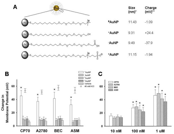
Summary of gold nanoparticles (AuNPs) effects on cellular membrane potential. AuNPs of different surface charges were generated by chemical modification of the terminal portion of the ligand bonded to the nanoparticle core. Four types of AuNPs were examined (A): neutral (0AuNP), positive (+AuNP), negative (−AuNP) and zwitterionic (±AuNP). The diameter (*) and surface charge (**) were measured by dynamic light scattering and zeta potential, respectively. Using the cell-permeant fluorescent membrane potential indicator RH414 and real-time fluorescence microscopy, membrane potential changes following exposure to AuNPs of different surface charges were measured for two ovarian cancer cell lines (CP70, A2780), human bronchial epithelial cells (BEC) and human airway smooth muscle (ASM) cells (also see supplemental Figure S1). +AuNPs (1.2 μM) produced rapid and significant membrane depolarization (panel B; bars; comparable to that induced by 40 mM KCl, diamonds). The extent of membrane potential change was dependent on +AuNPs concentration (C), with minimal changes at 10 nM. In comparison to +AuNPs, those with other charges had negligible effects on membrane potential in any cell type (B). Values are means ± SE. * indicates significant AuNP effect (p<0.05).
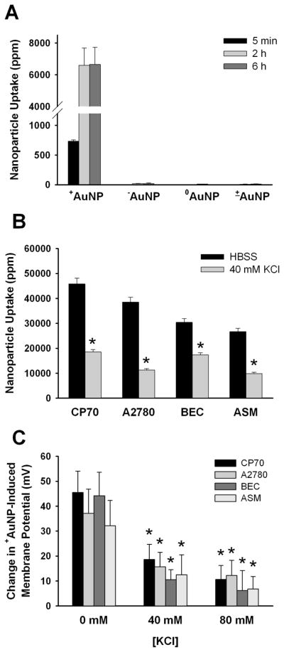
AuNP uptake and membrane potential. (A) In CP70 cells, only +AuNPs (0.4 μM) showed significant uptake, with substantial intracellular levels present at 5 min, followed by a higher level beyond 2h. (B) Prior exposure to KCl (inducing membrane depolarization) significantly (p<0.05) reduced the extent of uptake of +AuNPs (1.2 μM for 30 minutes treatment) in four different types of cells. (C) In all the cells investigated, prior exposure to KCl significantly blunted the extent of membrane depolarization subsequently induced by +AuNPs. Values are means ± SE. * indicates significant AuNP effect (p<0.05).
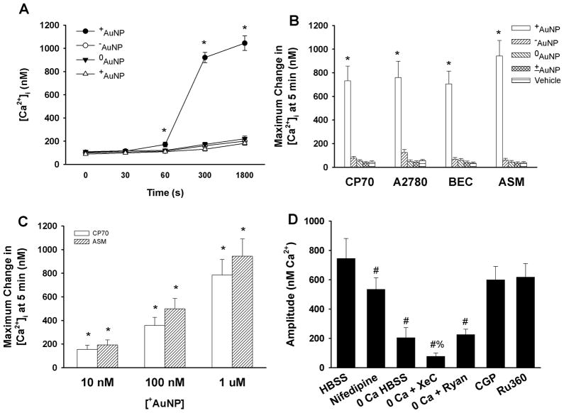
Summary of +AuNPs effects on intracellular Ca2+ ([Ca2+]i) levels. (A, B) In ovarian cancer cells (CP70, A2780) and airway cells (BEC, ASM) loaded with the ratiometric fluorescent Ca2+ indicator fura-2, +AuNPs (1.2 μM) produced substantial increases in [Ca2+]i levels that reached a maximum value in ~5 minutes and in some cell types decayed to a lower level above baseline (see supplemental Figure S2). In comparison, AuNPs with other charges had negligible effects on [Ca2+]i levels. The effect on [Ca2+]i levels was dependent on the concentration of +AuNPs (C), with even 10 nM AuNPs producing a substantial increase in [Ca2+]I (compared to small effects on membrane potential). In ovarian cancer CP70 cells, the role of specific [Ca2+]i regulatory mechanisms were examined by first inhibiting specific Ca2+ regulatory mechanisms, and then exposing cells to +AuNPs (1.2 μM) (D). Inhibition of plasma membrane Ca2+ influx via L-type Ca2+ channels resulted in significant reduction in positively charged AuNP effects on [Ca2+]i levels. AuNP effects were even more reduced in the absence of extracellular Ca2+ (0 Ca HBSS) suggesting that Ca2+ influx (partly via L-type channels) contributes to the observed change in [Ca2+]i levels with AuNPs. The remainder of the [Ca2+]i response appears to be derived from endoplasmic reticulum Ca2+ release (since the response persists in the absence of extracellular Ca2+) especially via IP3 receptor channels (evidenced by lack of AuNP effects when the channels are inhibited by Xestospongin C). Lack of effect of mitochondrial Ca2+ pathways (CGP 37,157 for mitochondrial Na+/Ca2+ exchange and Ru360 for mitochondrial Ca2+ uniporter) suggests that AuNPs may not be affecting mitochondria. Values are means ± SE. * indicates significant AuNP effect, and # indicates significant effect of 0 Ca HBSS, and % indicates significant effect of inhibitor (p<0.05).
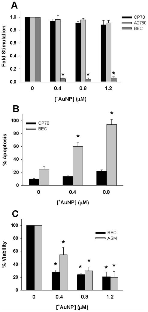
Effect of AuNPs on cellular proliferation vs. apoptosis. (A) Even brief (30 min) exposure of cells to +AuNPs substantially blunted the proliferation of human BECs, but did not affect the proliferation of ovarian cancer CP70 or A2780 cells. (B). Apoptosis of BEC by +AuNPs was concentration-dependent, and substantial. In contrast, some degree of apoptosis (< 10 %) of CP70 cells occurred only at a higher +AuNP concentration. (C) The viability of both BECs and ASM cells was substantially reduced by +AuNPs, with a concentration-dependence for ASM. * indicates significant effect (p<0.001)
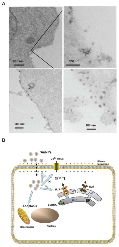
(A) Transmission electron microscopy of +AuNP (concentration = 0.4 μM) interactions with plasma membrane. Top panels demonstrate localization of +AuNPs within the plasma membrane (CP70 cells). Bottom panels demonstrate lack of plasma membrane disruption following +AuNP uptake (BEC cells). (B) Schematic of AuNP effects on cellular function. Based on our findings using +AuNPs and different types of cells, we propose that AuNPs are taken up intracellularly, based on membrane potential. Upon uptake, +AuNPs produce membrane depolarization, and increase [Ca2+]i by enhancing Ca2+ influx and inducing release of intracellular Ca2+ stores (e.g. via IP3 receptor channels of the endoplasmic reticulum; ER). These changes can result in increased apoptosis and decreased cellular proliferation, depending on cell type. Further modulation of apoptosis and proliferation may involve direct nanoparticle effects on intracellular signaling mechanisms.
Similar articles
-
Penetration of lipid membranes by gold nanoparticles: insights into cellular uptake, cytotoxicity, and their relationship.ACS Nano. 2010 Sep 28;4(9):5421-9. doi: 10.1021/nn1010792. ACS Nano. 2010. PMID: 20799717
-
Gold-Nanoparticle-Mediated Depolarization of Membrane Potential Is Dependent on Concentration and Tethering Distance from the Plasma Membrane.Bioconjug Chem. 2020 Mar 18;31(3):567-576. doi: 10.1021/acs.bioconjchem.9b00656. Epub 2020 Jan 14. Bioconjug Chem. 2020. PMID: 31894966
-
Gold nanoparticles with different charge and moiety induce differential cell response on mesenchymal stem cell osteogenesis.Biomaterials. 2015 Jun;54:226-36. doi: 10.1016/j.biomaterials.2015.03.001. Epub 2015 Apr 6. Biomaterials. 2015. PMID: 25858865
-
Gold Nanoparticles in Single-Cell Analysis for Surface Enhanced Raman Scattering.Molecules. 2016 Nov 25;21(12):1617. doi: 10.3390/molecules21121617. Molecules. 2016. PMID: 27897986 Free PMC article. Review.
-
Design and applications of gold nanoparticle conjugates by exploiting biomolecule-gold nanoparticle interactions.Nanoscale. 2013 Apr 7;5(7):2589-99. doi: 10.1039/c3nr33870c. Nanoscale. 2013. PMID: 23423633 Review.
Cited by
-
Development of a cationic polyethyleneimine-poly(lactic-co-glycolic acid) nanoparticle system for enhanced intracellular delivery of biologics.RSC Adv. 2023 Nov 17;13(48):33721-33735. doi: 10.1039/d3ra06050k. eCollection 2023 Nov 16. RSC Adv. 2023. PMID: 38020041 Free PMC article.
-
The membrane axis of Alzheimer's nanomedicine.Adv Nanobiomed Res. 2021 Jan;1(1):2000040. doi: 10.1002/anbr.202000040. Epub 2020 Nov 26. Adv Nanobiomed Res. 2021. PMID: 33748816 Free PMC article.
-
Biocompatible, Multi-Mode, Fluorescent, T2 MRI Contrast Magnetoelectric-Silica Nanoparticles (MagSiNs), for On-Demand Doxorubicin Delivery to Metastatic Cancer Cells.Pharmaceuticals (Basel). 2022 Sep 30;15(10):1216. doi: 10.3390/ph15101216. Pharmaceuticals (Basel). 2022. PMID: 36297329 Free PMC article.
-
Green pyomelanin-mediated synthesis of gold nanoparticles: modelling and design, physico-chemical and biological characteristics.Microb Cell Fact. 2019 Dec 3;18(1):210. doi: 10.1186/s12934-019-1254-2. Microb Cell Fact. 2019. PMID: 31796078 Free PMC article.
-
Role of Surface Hydrophobicity of Dicationic Amphiphile-Stabilized Gold Nanoparticles on A549 Lung Cancer Cells.ACS Omega. 2017 Jul 31;2(7):3527-3538. doi: 10.1021/acsomega.7b00353. Epub 2017 Jul 25. ACS Omega. 2017. PMID: 30023697 Free PMC article.
References
-
- Ferrari M. Nat Rev Cancer. 2005;5(3):161–71. - PubMed
-
- Burda C, Chen X, Narayanan R, El-Sayed MA. Chem Rev. 2005;105(4):1025–102. - PubMed
-
- Daniel MC, Astruc D. Chem Rev. 2004;104(1):293–346. - PubMed
-
- Patra CR, Bhattacharya R, Wang E, Katarya A, Lau JS, Dutta S, Muders M, Wang S, Buhrow SA, Safgren SL, Yaszemski MJ, Reid JM, Ames MM, Mukherjee P, Mukhopadhyay D. Cancer Res. 2008;68(6):1970–8. - PubMed
-
- Mirkin CA, Taton TA. Nature. 2000;405(6787):626–7. - PubMed
Publication types
MeSH terms
Substances
Grants and funding
LinkOut - more resources
Full Text Sources
Other Literature Sources
