Review
. 2010:79:707-35.
doi: 10.1146/annurev.biochem.77.060407.135452.

Physical mechanisms of signal integration by WASP family proteins

Affiliations
Review

Physical mechanisms of signal integration by WASP family proteins

Shae B Padrick et al. Annu Rev Biochem. 2010.

Abstract

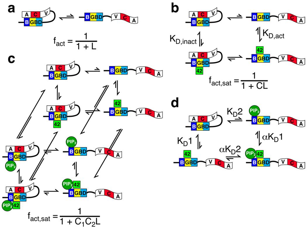
The proteins of the Wiskott-Aldrich syndrome protein (WASP) family are activators of the ubiquitous actin nucleation factor, the Arp2/3 complex. WASP family proteins contain a C-terminal VCA domain that binds and activates the Arp2/3 complex in response to numerous inputs, including Rho family GTPases, phosphoinositide lipids, SH3 domain-containing proteins, kinases, and phosphatases. In the archetypal members of the family, WASP and N-WASP, these signals are integrated through two levels of regulation, an allosteric autoinhibitory interaction, in which the VCA is sequestered from the Arp2/3 complex, and dimerization/oligomerization, in which multi-VCA complexes are better activators of the Arp2/3 complex than monomers. Here, we review the structural, biochemical, and biophysical details of these mechanisms and illustrate how they work together to control WASP activity in response to multiple inputs. These regulatory principles, derived from studies of WASP and N-WASP, are likely to apply broadly across the family.


Figures

Figure 1
Figure 1. The WASP family of proteins
(A) Domain structure of human WASP family proteins. Different domains are abbreviated as EVH1, Ena/VASP Homology domain 1; B, Basic region; GBD, GTPase Binding Domain; PRD, Proline Rich Domain; V, Verprolin homology domain (also WH2); C, Central hydrophobic region; A, Acidic region; SHD, Scar homology domain; WHD1, WASH homology domain 1; WHD2, WASH homology domain 2; NTD, WHAMM/JMY N-terminal homology domain; CC, Coiled Coil domain. In JMY, the NTD has an insertion of > 100 amino acids when compared to WHAMM. There are multiple closely related WASH proteins in humans, shown here is WASH1 (NCBI ref# NP_878908). (B) WASP has distinct binding sites for different ligands. Human WASP B-GBD sequence is shown with domains colored as in Figure 1. Secondary structure for the WASP GBD-C autoinhibited structure is shown (Figure 2c–e). Green bars indicate the binding sites for PIP2, Cdc42 and SH2 domain. Other annotations: dashed box, CRIB; Orange ovals, X-Linked neutropenia WASP mutations; green triangle, Tyrosine 291.
Figure 2
Figure 2. Structures of WASP
(A) The structure of WASP V/WH2-region in complex with actin. Actin is shown as a gray surface, WASP is shown as an orange ribbon. (B) Structure of the N-WASP EVH1:WIP(451–485) complex (structured WIP residues 454–481 shown). EVH1 domain is gold, proline motif (461DLPPPEPY468) and flanking regions of WIP are purple and blue, respectively. (C–E) Structure of autoinhibited WASP (GBD-C). (C) Structure of the GBD-C protein. The three layers of structure are colored yellow, blue (GBD) and red (C region of VCA), respectively. (D) Structure of the GBD-C protein, rotated 180° from panel C. GBD and C elements are shown in surface and ribbon representations, respectively. (E) Surface representation of GBD-C protein, rotated ~90° from panel C, showing Y291 phosphorylation site in green. (F–G) Structures of active WASP complexes. (F) Structure of the Cdc42:WASP GBD complex. Cdc42 is green ribbon, with GMPPNP in sticks. Ordered GBD residues (231–277) are shown in gold (CRIB motif and flanking sequence) or yellow (Layer 1 as in panel C) ribbon. (G) Structure of the WASP GBD:EspFU 1R complex. GBD is colored as in panel C and shown in surface representation. EspFU is shown as a green ribbon. (H) Structure of the WASP GBD-C:Wiskostatin complex, orientation and color as in panel E, with wiskostatin shown as van der Waals spheres.
Figure 3
Figure 3. Thermodynamic models of allostery in WASP
(A) Two-state MWC-based allosteric model for WASP regulation by control of access to the C-helix through association with the GBD. (B) Two-state allosteric model with one activator. (C) Two-state allosteric model with two activators. Equations describing the fraction of WASP in the active state, either free (fact) or in the presence of saturating ligand (fact,sat) are given below the images. L is defined as [WASPinactive]/[WASPactive]; C is the ratio of dissociation constants of a ligand for the active and inactive states, KD,act/KD,inact. (D) Simplified allosteric model for response to two activators. KD1 and KD2 are the dissociation constants of the two ligands for free WASP, α is the cooperativity constant between the ligands.
Figure 4
Figure 4. The hierarchical model of WASP regulation
WASP proteins are regulated hierarchically. An inner layer of allostery controls access of the VCA, and outer layer of dimerization/oligomerization controls affinity of the VCA for Arp2/3 complex. The two layers are thermodynamically coupled, so that binding of Arp2/3 complex to a VCA dimer will also shift the allosteric equilibrium toward the active state. Figure adapted by permission from Cell Press: Molecular Cell (22), copyright 2008.
Figure 5
Figure 5. Cooperative activation of N-WASP/WRC by higher order complex formation
Multi-protein complexes that bring two or more VCAs together. (A) Schematic illustration of the complex formed by N-WASP, Toca (dark red, with domains indicated), Cdc42 (labeled “42”), PIP2 (red circles) and Arp2/3 at membranes. (B) Schematic illustration of the complex formed by the WRC, IRSp53 (Blue with domains indicated), Rac, PIP3 (orange circles) and Arp2/3 at membranes. Inset shows organization of the WRC. (C) Schematic illustration of the complex formed by phosphorylated Cortactin, Nck, N-WASP and WIP. (D) Schematic illustration of the complex formed by phosphorylated Nephrin, Nck (purple ovals and green square) and N-WASP (domain colors as in Figure 1).

References

    1. Pollard TD. Regulation of actin filament assembly by Arp2/3 complex and formins. Annu Rev Biophys Biomol Struct. 2007;36:451–477. - PubMed
    1. Chhabra ES, Higgs HN. The many faces of actin: matching assembly factors with cellular structures. Nat Cell Biol. 2007;9:1110–1121. - PubMed
    1. Pollitt AY, Insall RH. WASP and SCAR/WAVE proteins: the drivers of actin assembly. J Cell Sci. 2009;122:2575–2578. - PMC - PubMed
    1. Kurisu S, Takenawa T. The WASP and WAVE family proteins. Genome Biol. 2009;10:226. - PMC - PubMed
    1. Stradal TE, Scita G. Protein complexes regulating Arp2/3-mediated actin assembly. Curr Opin Cell Biol. 2006;18:4–10. - PubMed

Publication types

MeSH terms

Substances

LinkOut - more resources