Review
. 2010:47:85-98.
doi: 10.1042/bse0470085.

Mitochondrial fission and fusion

Affiliations
Review

Mitochondrial fission and fusion

Iain Scott et al. Essays Biochem. 2010.

Abstract

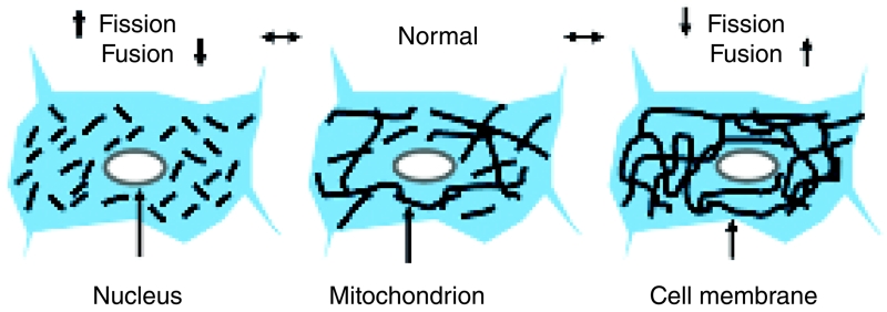
Mitochondria are highly dynamic cellular organelles, with the ability to change size, shape and position over the course of a few seconds. Many of these changes are related to the ability of mitochondria to undergo the highly co-ordinated processes of fission (division of a single organelle into two or more independent structures) or fusion (the opposing reaction). These actions occur simultaneously and continuously in many cell types, and the balance between them regulates the overall morphology of mitochondria within any given cell. Fission and fusion are active processes which require many specialized proteins, including mechanical enzymes that physically alter mitochondrial membranes, and adaptor proteins that regulate the interaction of these mechanical proteins with organelles. Although not fully understood, alterations in mitochondrial morphology appear to be involved in several activities that are crucial to the health of cells. In the present chapter we discuss the mechanisms behind mitochondrial fission and fusion, and discuss the implications of changes in organelle morphology during the life of a cell.


Figures

Figure 1
Figure 1. Balance of fission and fusion dynamically regulates mitochondrial morphology
The overall morphology of mitochondria within any given cell is dynamically controlled by a balance between organelle fission and fusion. In many cell types, mitochondria form a network of interconnected tubules, along with multiple independent organelles (centre). Continual fission and fusion events lead to rapid changes in morphology in discrete areas; however, when these two processes are balanced, the overall mitochondrial morphology observed appears relatively constant. When the balance between fission and fusion is disrupted, caused by a relative increase or decrease in either process, the overall mitochondrial morphology can change markedly. In fusion-deficient cells (left-hand side), continued fission leads to the break-up of the mitochondrial network, leading to an increase in smaller independent organelles. Conversely, in fission-deficient cells (right-hand side), the relative increase in fusion events leads to a highly interconnected network of mitochondrial tubules.
Figure 2
Figure 2. Mitochondrial fission in mammalian cells
The two major proteins involved in mammalian mitochondrial fission are Fis1 (black triangle) and Drp1 (grey oval), and loss of either protein limits fission. Fis1 is an adaptor protein located in the OMM, where it is anchored by a C-terminal transmembrane domain. The remainder of Fis1 consists of protein–protein interaction sites which protrude into the cytosol. The majority of Drp1 is localized in the cytosol, from where it shuttles back and forth to the OMM during fission events. Drp1 interacts with Fis1 at fission sites, where it is proposed to form a collar that encircles the mitochondrion. The Drp1 collar tightens around the mitochondrion, and this constriction leads to a severing of the OMM and fission into two independent organelles. What regulates the site of fission, and how fission of the IMM is carried out, is currently unknown. Mff is a second OMM protein, which is anchored at its C-terminus like Fis1. Loss of Mff blocks mitochondrial fission; however, its mode of action, and whether it interacts with Drp1 to aid fission, is currently unknown. IMS, intermembrane space.
Figure 3
Figure 3. Mitochondrial fusion in mammalian cells
The major fusion proteins of mammalian mitochondria are Mfn1 and Mfn2 (black symbol) and OPA1 (grey symbol). Mfn proteins are localized to the OMM and aid the tethering of two discrete mitochondria at the early stages of fusion, through homotypic coiled-coil protein-interaction domains. These proteins are also hypothesized to provide the mechanical forces involved in outer membrane fusion. mitoPLD (white symbol) is also localized to the OMM, with its catalytic domain facing the cytosol. Topological and enzymatic studies suggest that it may insert into the membranes of neighbouring mitochondria, changing the properties of the membrane lipids to aid fusion. OPA1 is localized in the IMM and intermembrane space, and co-ordinates the fusion of the IMM. OPA1 is processed into several different cleavage isoforms by mitochondrial proteases, such as PARL (blue symbol), with each variant proposed to have different roles in fusion events.

Similar articles

Cited by

References

    1. Kennedy EP, Lehninger AL. Oxidation of fatty acids and tricarboxylic acid cycle intermediates by isolated rat liver mitochondria. J. Biol. Chem. 1949;179:957–972. - PubMed
    1. Lewis MR, Lewis WH. Mitochondria in tissue cultures. Science. 1914;39:330–333. - PubMed
    1. Suen D-F, Norris KL, Youle RJ. Mitochondrial dynamics and apoptosis. Genes Dev. 2008;22:1577–1590. - PMC - PubMed
    1. Sesaki H, Jensen RE. Division versus fusion: Dnm1p and Fzo1p antagonistically regulate mitochondrial shape. J. Cell Biol. 1999;149:699–706. - PMC - PubMed
    1. Zimmer C. On the origin of eukaryotes. Science. 2009;325:666–668. - PubMed

Publication types