Review
. 2010 May 28;17(5):421-33.
doi: 10.1016/j.chembiol.2010.04.012.

DNA topoisomerases and their poisoning by anticancer and antibacterial drugs

Affiliations
Review

DNA topoisomerases and their poisoning by anticancer and antibacterial drugs

Yves Pommier et al. Chem Biol. .

Abstract

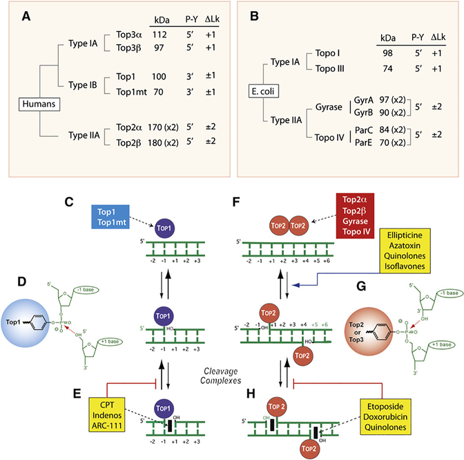
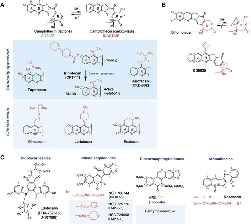
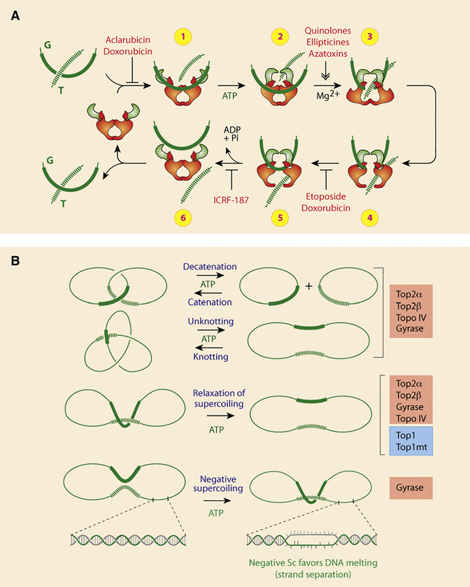
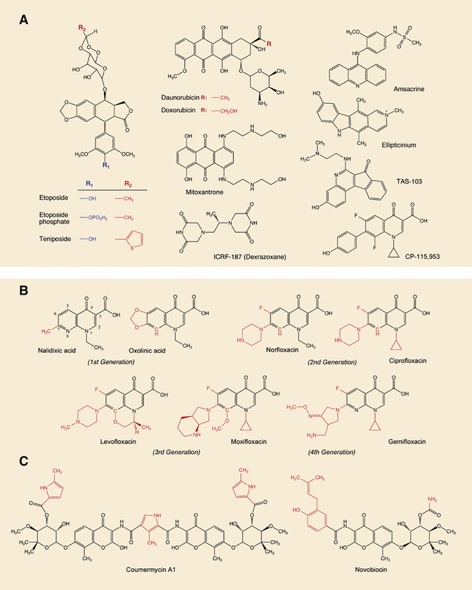
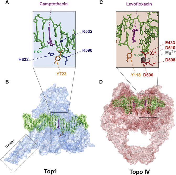
DNA topoisomerases are the targets of important anticancer and antibacterial drugs. Camptothecins and novel noncamptothecins in clinical development (indenoisoquinolines and ARC-111) target eukaryotic type IB topoisomerases (Top1), whereas human type IIA topoisomerases (Top2alpha and Top2beta) are the targets of the widely used anticancer agents etoposide, anthracyclines (doxorubicin, daunorubicin), and mitoxantrone. Bacterial type II topoisomerases (gyrase and Topo IV) are the targets of quinolones and aminocoumarin antibiotics. This review focuses on the molecular and biochemical characteristics of topoisomerases and their inhibitors. We also discuss the common mechanism of action of topoisomerase poisons by interfacial inhibition and trapping of topoisomerase cleavage complexes.


Figures

Figure 1.
Figure 1.. Overview of DNA Topoisomerases
(A) Classification of human DNA topoisomerases. kDa: molecular masses calculated from polypeptide composition. (x2): dimer. Type IB are the only enzymes that form cleavage complexes (cc) with 3′-phosphotyrosyl (3′-P-Y) intermediates. ΔLk: linking number change produced by each catalytic cycle. Type I enzymes change Lk in steps of one and type II in steps of two as they cleave one and both strands of DNA, respectively. Type IA enzymes are the only enzymes that relax only negative but not positive supercoiling (ΔLk = +1). (B) Classification of E. coli DNA topoisomerases. (C) Noncovalent binding of type IB enzymes. (D) Scheme of the 3′-phosphotyrosine covalent bond in the Top1cc. The arrow indicates the reversible (religation) reaction, which is favored under normal conditions. (E) Trapping of the cleavage complex by camptothecin (CPT) and the Top1 inhibitors (see Figure 3). (F) Noncovalent binding of type IIA enzyme homodimers (A2B2 and C2E2 in the case of gyrase and Topo IV, respectively). (G) Scheme of the 5′-phosphotyrosine covalent bond in the Top2cc. The arrow indicates the reversible (religation) reaction, which is favored under normal conditions. Note the four base pair stagger with 5′-overhang on opposite strands characteristic of all type IIA enzymes. (H) Trapping of the cleavage complex by etoposide, doxorubicin, or quinolones (see Figure 3). Note that some Top2 poisons increase the steady-state levels of Top2cc by increasing cleavage (ellipticines, azatoxin, quinolones, and isoflavones).
Figure 2.
Figure 2.. Overview of Type IB Topoisomerases and Relaxation by Nicking-Closing Activity
(A) Schematic representation of the two human type IB enzymes. Top1mt has a much shorter N-terminal segment consisting primarily of a mitochondrial targeting sequence (MTS) and lacking the nuclear localization sequences (NLS) of Top1 (nuclear). The catalytic homologous residues are indicated with their position. (B) Schematic representation of the poxvirus (variola and vaccinia) Top1s and comparison with the recently discovered bacterial Top1B (Pseudomonas aeroginosa). (C) Top1-mediated DNA relaxation by controlled rotation. By contrast to type IA or II enzymes, this reaction does not require an energy cofactor or divalent metal. Top1 tends to bind DNA crossovers (supercoils) and nicks DNA by transesterification (see Figure 1D). The enzyme then allows the DNA to swivel by controlled rotation (Koster et al., 2005; Stivers et al., 1997). Upon DNA realignment by base pairing and stacking across the nick, the DNA 5′-hydroxyl end (OH in lower panels) removes the tyrosyl linkage by reverse transesterification (see Figure 1D).
Figure 3.
Figure 3.. Top1 Inhibitors
(A) Camptothecin and its clinical derivatives. The facile and reversible opening of the α-hydroxylactone E ring of camptothecin is shown at the top. Topotecan and irinotecan are the two FDA-approved camptothecins. Irinotecan is a prodrug; its active metabolite is SN-38. Belotecan is approved in South Korea. (B) Synthetic E-ring-modified camptothecin derivatives. Whereas diflomotecan (a homocamptothecin) can still be converted irreversibly to a carboxylate, the α-keto derivative S39625 is chemically stable while still being a potent Top1 inhibitor. (C) Noncamptothecins. The indenoisoquinolines and one dibenzonaphthyridinone are beginning clinical trials.
Figure 4.
Figure 4.. Structural Overview of the Type IIA Topoisomerases Targeted by Anticancer and Antibacterial Drugs
(A) Schematic representation of the human Top2 enzymes Top2α and β. Note the high similarity of their ATPase and cleavage/religation domains, whereas their N- and C-terminal regions are more divergent. Conserved catalytic residues are indicated including the catalytic tyrosines Y805 and Y821 in Top2α and β, respectively. (B) Schematic representation of the bacterial Top2 enzymes gyrase and Topo IV. Gyrase is a heterotetramer encoded by two genes: GyrA and GyrB (A2B2; see Figure 1B). Topo IV is also a heterotetramer encoded by two genes: ParC and ParE (C2E2; see Figure 1B). Conserved catalytic residues are indicated including the catalytic tyrosines Y122 andY120 ingyrase and Topo IV, respectively. (C) Conserved interaction of type IIA enzymes with DNA by their TOPRIM motifs (alignment at left). The right scheme represents the two-metal-binding model between the TOPRIM motif and DNA (modified from Deweese et al., 2008).
Figure 5.
Figure 5.. Catalytic Cycle and Reactions Carried Out by Type IIA Topoisomerases
(A) Schematic representation of the catalytic cycle for human Top2 enzymes (see text for details). Note that doxorubicin can block the catalytic cycle at two different steps. At low concentration (<1 μM), doxorubicin acts like etoposide (VP-16) by blocking DNA religation (between steps 4 and 5). At higher concentrations (>10 μM), doxorubicin acts like aclarubicin by interfering with Top2 binding to DNA (before step 1). ICRF-187 blocks ATP hydrolysis and inhibits reopening of the ATPase domain (between steps 5 and 6), thereby trapping topological complexes with DNA inside the enzyme. (B) Reactions catalyzed by type IIA topoisomerase. Note that Top1 can only catalyze supercoiling relaxation. Also, in bacteria, Topo IV acts preferentially as the decatenation enzyme, whereas gyrase acts preferentially in removing positive supercoiling and selectively in generating negative supercoiling.
Figure 6.
Figure 6.. Inhibitors of Type IIA Topoisomerases
(A) Inhibitors of eukaryotic Top2 enzymes. All drugs except ICRF-187 trap Top2 cleavage complexes and generate DNA breaks (see Figures 1F–1H). ICRF-187 is a catalytic inhibitor. (B) Gyrase and Topo IV inhibitors from the quinolone family. All drugs trap cleavage complexes. Note that the quinolone CP-115,953 traps both eukaryotic and prokaryotic enzymes, which is not the case in the other quinolones shown in this panel. Also note that TAS-103 is a dual inhibitor of Top1 and Top2 (Wilson Byl et al., 1999). (C) Gyrase catalytic inhibitors.
Figure 7.
Figure 7.. Interfacial Inhibition by Topoisomerase Inhibitors
(A) Intercalation of camptothecin in the DNA break between the base pairs flanking the Top1 cleavage complex (positions −1 and +1; see Figures 1C–1E). The basic residues that catalyze the nucleophilic attack of DNA by the catalytic tyrosine (Y743 in gold) are shown in blue (see Figure 2A). The Top1 amino acid residues that make hydrogen-bond interactions with the camptothecin E ring are not shown (see Figure 4 in Marchand et al., 2006). (B) Overview of the Top1-DNA cleavage complex.Top1 is in blue, DNA is in green, and camptothecin is in purple (Ioanoviciu et al., 2005; Marchand et al., 2006) (Protein Data Bank ID code 1T8I). (C) Intercalation of levofloxacin in the DNA break between the base pairs that flank the Topo IV cleavage complex (−1 and +1; see Figure 1). The acidic residues that coordinate the Mg2+ (see Figures 4B and 4C) are shown in red and the catalytic tyrosine (Y118) in gold. (D) Overview of the Topo IV-DNA cleavage complex. Topo IV is in red, the DNA is in green, and the levofloxacin molecules in both cleavage sites are in purple (Laponogov et al., 2009) (Protein Data Bank ID code 3K9F).

Similar articles

Cited by

References

    1. Azarova AM, Lyu YL, Lin CP, Tsai YC, Lau JY, Wang JC, and Liu LF (2007). Roles of DNA topoisomerase II isozymes in chemotherapy and secondary malignancies. Proc. Natl. Acad. Sci. USA 104, 11014–11019. - PMC - PubMed
    1. Bertrand R, O’Connor P, Kerrigan D, and Pommier Y (1992). Sequential administration of camptothecin and etoposide circumvents the antagonistic cytotoxicity of simultaneous drug administration in slowly growing human colon carcinoma HT-29 cells. Eur. J. Cancer 28A, 743–748. - PubMed
    1. Black MT, and Coleman K (2009). New inhibitors of bacterial topoisomerase GyrA/ParC subunits. Curr. Opin. Investig. Drugs 10, 804–810. - PubMed
    1. Bradbury BJ, and Pucci MJ (2008). Recent advances in bacterial topoisomerase inhibitors. Curr. Opin. Pharmacol. 8, 574–581. - PubMed
    1. Brangi M, Litman T, Ciotti M, Nishiyama K, Kohlhagen G, Takimoto C, Robey R, Pommier Y, Fojo T, and Bates SE (1999). Camptothecin resistance: role of the ATP-binding cassette (ABC), mitoxantrone-resistance half-transporter (MXR), and potential for glucuronidation in MXR-expressing cells. Cancer Res. 59, 5938–5946. - PubMed

Publication types

MeSH terms