. 2010 Jun 22;107(25):11352-7.
doi: 10.1073/pnas.1000142107. Epub 2010 Jun 3.

Structure of the full-length Shaker potassium channel Kv1.2 by normal-mode-based X-ray crystallographic refinement

Affiliations

Structure of the full-length Shaker potassium channel Kv1.2 by normal-mode-based X-ray crystallographic refinement

Xiaorui Chen et al. Proc Natl Acad Sci U S A. .

Abstract

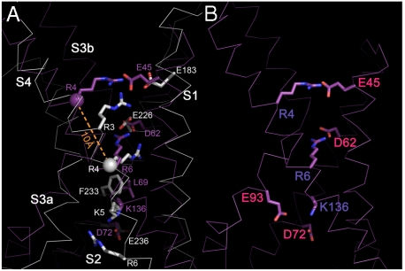
Voltage-dependent potassium channels (Kv) are homotetramers composed of four voltage sensors and one pore domain. Because of high-level structural flexibility, the first mammalian Kv structure, Kv1.2 at 2.9 A, has about 37% molecular mass of the transmembrane portion not resolved. In this study, by applying a novel normal-mode-based X-ray crystallographic refinement method to the original diffraction data and structural model, we established the structure of full-length Kv1.2 in its native form. This structure offers mechanistic insights into voltage sensing. Particularly, it shows a hydrophobic layer of about 10 A at the midpoint of the membrane bilayer, which is likely the molecular basis for the observed "focused electric field" of Kv1.2 between the internal and external solutions. This work also demonstrated the potential of the refinement method in bringing up large chunks of missing densities, thus beneficial to structural refinement of many difficult systems.


Conflict of interest statement

The authors declare no conflict of interest.

Figures

Fig. 1.
Fig. 1.
Overall structural improvements. (A) The original Kv1.2 structure (PDB ID code 2A79). (B) The final Kv1.2 structure after normal-mode refinement and multicrystal averaging. In both A and B, red represents the regions newly added, including the S1-S2 linker, S2-S3 linker, and S3-S4 linker; green highlights the T1-S1 linker where large structural adjustments were made and register was determined; cyan suggests the regions (the S1 helix and S3 helix) the registers of which were undetermined in the original Kv1.2 structure but have been fixed in the normal-mode-refined structure; and yellow indicates the rebuilding of the missing side chains on S2 helix, S4 helix, and the linker between S5 and the pore helix. (CF′′) Examples of structural improvements in normal-mode refinement comparing the original model (CF), the normal-mode model (C′–F′), and the final model after normal-mode refinement and multicrystal averaging (C′′–F′′), superimposed with their respective electron density maps. Shown are the T1-S1 linker region (C, C′, C′′), the S1-S2 linker (D, D′, D′′), the S2-S3 linker (E, E′, E′′), and the S3-S4 linker (F, F′, F′′). All maps were 2Fo - Fc maps contoured at 0.8σ.
Fig. 2.
Fig. 2.
Comparison of the transmembrane portions of the final Kv1.2 structure and the paddle-chimera structure. (A) Ribbon diagram of the paddle chimera structure with the chimera paddle region colored in red. (B) Ribbon diagram of the final Kv1.2 structure. The glycosylation site Asn207 (mutated from Gln) in A and B is highlighted as yellow stick. (C) Stereo view of the superposition of transmembrane portion of the final Kv1.2 structure (White) and the paddle-chimera structure (Cyan). Potassium ions are shown as purple spheres in AC. (D) The voltage sensors of the final Kv1.2 structure (White). (E) The voltage sensors of the paddle-chimera structure (Cyan). Key residues were shown as sticks for positive gating charges (Blue) and their interacting negatively charged residues (Red).
Fig. 3.
Fig. 3.
Comparison of the final Kv1.2 structure and the KvAP structure (PDB ID code 1ORS). (A) The superposition of the voltage sensors of the final Kv1.2 structure (White) and the KvAP structure (Purple). The residues R4 in S4 helix were highlighted with a sphere at its Cα atom in two structures, and the distance between them was indicated as a dashed line in orange. (B) The charged residues of the voltage sensor in KvAP structure. Key residues were shown as sticks for positive gating charges (Blue) and their interacting negatively charged residues (Red).
Fig. 4.
Fig. 4.
A hydrophobic layer in the voltage sensor of the final Kv1.2 structure. Hydrophobic residues are colored in magenta and hydrophilic residues in cyan. Transparent planes were drawn at both the upper and lower boundaries of the hydrophobic layer to highlight its thickness.

Similar articles

Cited by

References

    1. Hille B. Ion Channels of Excitable Membranes. 3rd Ed. Sunderland: Sinauer Associates; 2001.
    1. Yellen G. The moving parts of voltage-gated ion channels. Q Rev Biophys. 1998;31(3):239–295. - PubMed
    1. Tombola F, Pathak MM, Isacoff EY. How far will you go to sense voltage? Neuron. 2005;48(5):719–725. - PubMed
    1. Tombola F, Pathak MM, Isacoff EY. How does voltage open an ion channel? Annu Rev Cell Dev Biol. 2006;22:23–52. - PubMed
    1. MacKinnon R. Potassium channels. FEBS Lett. 2003;555(1):62–65. - PubMed

Publication types

Associated data