. 2010 Jun 15;107(24):10978-83.
doi: 10.1073/pnas.1006545107. Epub 2010 Jun 1.

Diabetogenic T cells recognize insulin bound to IAg7 in an unexpected, weakly binding register

Affiliations

Diabetogenic T cells recognize insulin bound to IAg7 in an unexpected, weakly binding register

Brian D Stadinski et al. Proc Natl Acad Sci U S A. .

Abstract

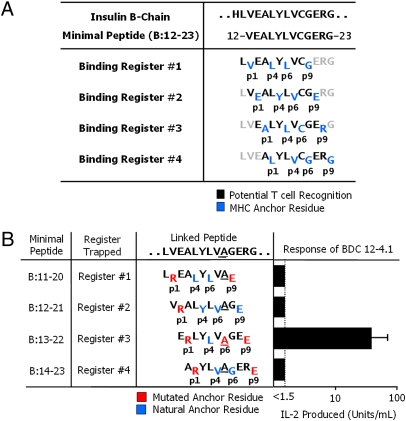
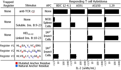
A peptide derived from the insulin B chain contains a major epitope for diabetogenic CD4(+) T cells in the NOD mouse model of type 1 diabetes (T1D). This peptide can fill the binding groove of the NOD MHCII molecule, IA(g7), in a number of ways or "registers." We show here that a diverse set of NOD anti-insulin T cells all recognize this peptide bound in the same register. Surprisingly, this register results in the poorest binding of peptide to IA(g7). The poor binding is due to an incompatibility between the p9 amino acid of the peptide and the unique IA(g7) p9 pocket polymorphisms that are strongly associated with susceptibility to T1D. Our findings suggest that the association of autoimmunity with particular MHCII alleles may be do to poorer, rather than more favorable, binding of the critical self-epitopes, allowing T-cell escape from thymic deletion.


Conflict of interest statement

The authors declare no conflict of interest.

Figures

Fig. 1.
Fig. 1.
The BDC 12-4.1 T cell responds to register 3 of the insulin B:12–23 peptide. (A) Four possible IAg7-binding registers for the mouse insulin B-chain peptide, B:12–23. Anchor residues are shown in blue, and black residues show the amino acids that potentially contact the T-cell antigen receptor. (B) Minimal 10-mer linked peptide versions of the insulin B:12–23(19A) epitope. Each minimal peptide was trapped in a single binding register by a p1Arg and a p9Glu. The substitution of Ala for Cys at B:19 of the peptide is underlined. Red amino acids are the substituted anchor residues. Blue amino acids are the naturally occurring anchor residues. Black amino acids are the possible T-cell epitope residues. Also shown is the stimulation of the BDC 12-4.1 T-cell hybridoma with each of the individual IAg7-linked peptide complexes expressed on the surface of ICAM/B7+ insect cells, assessed by the production of IL-2. Results are the average ± SEM of triplicate wells from five independent experiments.
Fig. 2.
Fig. 2.
Four independent insulin B-chain responsive T cells recognize the peptide bound to IAg7 in register 3. Triplicate culture wells of the BDC 12-4.1, AS91, AS150, and I.29 insulin B-chain responsive CD4+ T-cell hybridomas were stimulated with an immobilized anti-Cβ Mab (five experiments), NOD spleen cells with or without 50 μg/mL of soluble insulin B:9–23 peptide (two experiments), ICAM/B7+-infected insect cells expressing IAg7 linked to the control hen egg lysozyme (HEL) peptide or to the unmutated B:12–23 peptide (three experiments), or ICAM/B7+-infected insect cells expressing IAg7 linked to each of three trapped versions (Fig. S6) of the insulin B:10–23 peptide (three experiments). The responses of the hybridomas were assessed by IL-2 production, and the average response and SEM is shown.
Fig. 3.
Fig. 3.
The p9 Arg-to-Glu mutation dramatically improves the binding of a register 3 peptide to IAg7. (A) Various concentrations of either unmutated (blue) or p1Arg/p9Glu (red) versions of the minimal nonamer peptides for registers 1, 2, and 3 of the B:12–23(19A) peptide were compared with a HEL peptide (positive control, black squares) and an MCC peptide (negative control, black diamonds) for the ability to inhibit the binding of a biotinylated HEL peptide to soluble IAg7. The panels show the percent of biotinylated HEL peptide remaining bound to IAg7 vs. the dose of inhibitor. The results are for a representative experiment. (B) The experiment described in A was performed three times. In each experiment the inhibition curve for each peptide was compared with the curve obtained with the HEL peptide to determine its IAg7 binding ability relative to HEL expressed as a percent. The results presented are the average value for the three experiments with the SEM. Blue bars, unmutated peptides; red bars, p1Arg/p9Glu peptides; black bars, control peptides. (C) The relative ability of the unmutated register-3 nonamer peptide to bind to IAg7 (blue bar) was compared with register-3 peptides bearing p1Arg and/or p9Glu (red bars) and to the HEL and MCC control peptides (black bars), as in A and B. The results are the average ± SEM of three experiments.
Fig. 4.
Fig. 4.
Confirmation by disulfide bond formation that the insulin peptide is trapped in register 3. The structure of IAg7 bound to a GAD peptide (PDB ID code 1ES0) (11) was used to model (Swiss PDB Viewer) (39) cysteines replacing the amino acids either at peptide p6 and IAg7 α62Asn (A) or at peptide p11 and IAg7 α72Ile (B). In both cases, the distance and orientation of the cysteines predict the formation of a disulfide bond. (C) The stimulating register 3 complex in Fig. 2 was altered to remove its baculovirus gp64 transmembrane anchor. The complex was mutated to introduce a Cys into the IAg7 α chain at either 62Asn or 72Ile. The peptide-encoding region of the α72Ile>Cys complex was further mutated to convert the Cys at p6 to Ala and the Ala at p11 to Cys (Fig. S6). Infected insect cells were infected with baculovirus encoding for each complex and the soluble IAg7 purified from the culture supernatant. The soluble IAg7 complexes were analyzed by SDS/PAGE under nonreducing and reducing conditions. The gels were stained with Coomassie blue. (D) The baculovirus gp64 transmembrane anchor was restored to each complex shown in C, allowing expression via baculovirus on ICAM/B7+ insect cells, which were used to stimulate the four T-cell hybridomas. The responses were assessed by IL-2 production, and the average of three experiments with triplicate wells are shown with the SEM.

References

    1. Erlich H, et al. Type 1 Diabetes Genetics Consortium HLA DR-DQ haplotypes and genotypes and type 1 diabetes risk: Analysis of the type 1 diabetes genetics consortium families. Diabetes. 2008;57:1084–1092. - PMC - PubMed
    1. Prochazka M, Leiter EH, Serreze DV, Coleman DL. Three recessive loci required for insulin-dependent diabetes in nonobese diabetic mice. Science. 1987;237:286–289. - PubMed
    1. Todd JA, Bell JI, McDevitt HO. HLA-DQ beta gene contributes to susceptibility and resistance to insulin-dependent diabetes mellitus. Nature. 1987;329:599–604. - PubMed
    1. Suri A, Walters JJ, Gross ML, Unanue ER. Natural peptides selected by diabetogenic DQ8 and murine I-A(g7) molecules show common sequence specificity. J Clin Invest. 2005;115:2268–2276. - PMC - PubMed
    1. Reich EP, et al. Self peptides isolated from MHC glycoproteins of non-obese diabetic mice. J Immunol. 1994;152:2279–2288. - PubMed

Publication types

MeSH terms