. 2010 Jun 15;107(24):10990-5.
doi: 10.1073/pnas.1004153107. Epub 2010 Jun 1.

Crystal structure of soluble MD-1 and its interaction with lipid IVa

Affiliations

Crystal structure of soluble MD-1 and its interaction with lipid IVa

Sung-il Yoon et al. Proc Natl Acad Sci U S A. .

Abstract

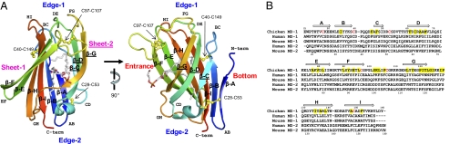
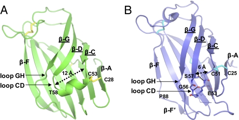
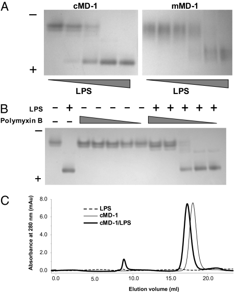
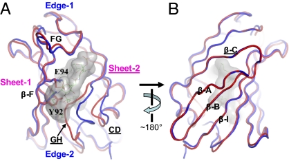
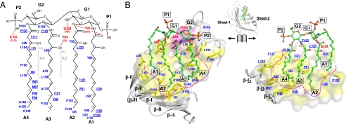
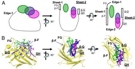
Lipopolysaccharide (LPS) of Gram-negative bacteria is a common pathogen-associated molecular pattern (PAMP) that induces potent innate immune responses. The host immune response against LPS is triggered by myeloid differentiation factor 2 (MD-2) in association with Toll-like receptor 4 (TLR4) on the cell surface. The MD-2/TLR4-mediated LPS response is regulated by the evolutionarily related complex of MD-1 and Toll-like receptor homolog RP105. Here, we report crystallographic and biophysical data that demonstrate a previously unidentified direct interaction of MD-1 with LPS. The crystal structure of chicken MD-1 (cMD-1) at 2.0 A resolution exhibits a beta-cup-like fold, similar to MD-2, that encloses a hydrophobic cavity between the two beta-sheets. A lipid-like moiety was observed inside the cavity, suggesting the possibility of a direct MD-1/LPS interaction. LPS was subsequently identified as an MD-1 ligand by native gel electrophoresis and gel filtration analyses. The crystal structure of cMD-1 with lipid IVa, an LPS precursor, at 2.4 A resolution revealed that the lipid inserts into the deep hydrophobic cavity of the beta-cup-like structure, but with some important differences compared with MD-2. These findings suggest that soluble MD-1 alone, in addition to its complex with RP105, can regulate host LPS sensitivity.


Conflict of interest statement

The authors declare no conflict of interest.

Figures

Fig. 1.
Fig. 1.
cMD-1 structure and sequence. (A) Overall structure of cMD-1 and its hydrophobic cavity. The cMD-1 structure is shown in ribbons that are colored from N terminus (blue) to C terminus (red). Labels for β-strands in sheet-2 are underlined. Disulfide bonds are shown in yellow ball-and-stick models, and a putative PGT molecule inside the MD-1 cavity is shown in sticks (carbon, gray; oxygen, red; phosphorus, orange). (B) MD-1 and MD-2 sequence alignment. cMD-1 cysteine residues that form disulfide bonds are colored in red, and cMD-1 residues that make contacts with lipid IVa are highlighted in yellow. cMD-1 β-strands are shown as arrows above the cMD-1 sequence.
Fig. 2.
Fig. 2.
Structural comparison between cMD-1 (A) and hMD-2 (B; PDB ID code 2e56). Regions that differ between MD-1 and MD-2 are highlighted.
Fig. 3.
Fig. 3.
Interaction of MD-1 with LPS. (A) Native PAGE of cMD-1/LPS (Left) and mMD-1/LPS (Right) mixtures. (B) Native PAGE shows competition of cMD-1 with polymyxin B for LPS. (C) Gel filtration analysis of cMD-1, LPS, and their mixture.
Fig. 4.
Fig. 4.
Conformational changes of cMD-1 upon lipid IVa binding. (A) Cavity entrance view. cMD-1PGT and cMD-1lipid IVa are displayed by red and blue coils, respectively. Lipid IVa bound to cMD-1 is shown as surface representation (gray) and also depicted in lines. (B) The view from the cavity bottom.
Fig. 5.
Fig. 5.
Interaction of cMD-1 with lipid IVa observed in the cMD-1/lipid IVa structure. (A) Schematic diagram of lipid IVa and interacting cMD-1 residues. Lipid IVa atoms that were not observed in the cMD-1/lipid IVa structure are represented by broken lines. Two extra lipid A acyl chains that are not present in lipid IVa are shown by light gray lines. cMD-1 residues that make H bonds (red dashed lines) with lipid IVa are labeled in red, and the remaining interacting residues are in blue. Residues of the sheet-2 side are underlined to discriminate those from sheet-1. (B) Open-book view of the cMD-1/lipid IVa interaction. To achieve the view, the cMD-1 structure (gray ribbons) shown in the middle was split between sheets-1 and -2, and then the sheet-1 (Left) and sheet-2 (Right) sides were rotated by 90° in the left and right directions, respectively, along the vertical axis. Lipid IVa is depicted in a ball-and-stick model (green). cMD-1 residues that form the internal cavity are color coded in surface representation (polar residues H/N/Q/S/T/C, gray; charged residues K/R/D/E, magenta; aliphatic and aromatic residues A/G/I/L/M/W/F/W/Y, yellow). cMD-1 residues that interact with lipid IVa are shown in sticks. The internal surface of the cavity is outlined in magenta broken lines.
Fig. 6.
Fig. 6.
Comparison of lipid IVacMD-1, lipid IVahMD-2 (PDB ID code 2e59), and LPShMD-2/hsTLR4 (PDB ID code 3fxi) structures. For clarity, only the lipid A portion of LPS is shown. (A) Schematic diagram of the relative ligand disposition in the hydrophobic cavities of MD-1 and MD-2. The phosphosugar backbone of ligand is represented by a circle (lipid IVacMD-1, green; lipid IVahMD-2, magenta; LPShMD-2/hsTLR4, blue). The internal surface of the cavity is schematically shown by broken lines. (B) The relative location and disposition of lipid IVacMD-1 (green), lipid IVahMD-2 (magenta), and LPShMD-2/hsTLR4 (blue) as observed in the superimposed MD-1 and MD-2 structures (yellow).

Similar articles

Cited by

References

    1. Raetz CR, Whitfield C. Lipopolysaccharide endotoxins. Annu Rev Biochem. 2002;71:635–700. - PMC - PubMed
    1. Gangloff M, Gay NJ. MD-2: The Toll ‘gatekeeper’ in endotoxin signalling. Trends Biochem Sci. 2004;29:294–300. - PubMed
    1. Martin GS, Mannino DM, Eaton S, Moss M. The epidemiology of sepsis in the United States from 1979 through 2000. N Engl J Med. 2003;348:1546–1554. - PubMed
    1. Beutler B, Rietschel ET. Innate immune sensing and its roots: The story of endotoxin. Nat Rev Immunol. 2003;3:169–176. - PubMed
    1. Shimazu R, et al. MD-2, a molecule that confers lipopolysaccharide responsiveness on Toll-like receptor 4. J Exp Med. 1999;189:1777–1782. - PMC - PubMed

Publication types

MeSH terms

LinkOut - more resources