. 2010 Jun 22;107(25):11483-8.
doi: 10.1073/pnas.1004600107. Epub 2010 Jun 2.

Structure and function of broadly reactive antibody PG16 reveal an H3 subdomain that mediates potent neutralization of HIV-1

Affiliations

Structure and function of broadly reactive antibody PG16 reveal an H3 subdomain that mediates potent neutralization of HIV-1

Robert Pejchal et al. Proc Natl Acad Sci U S A. .

Abstract

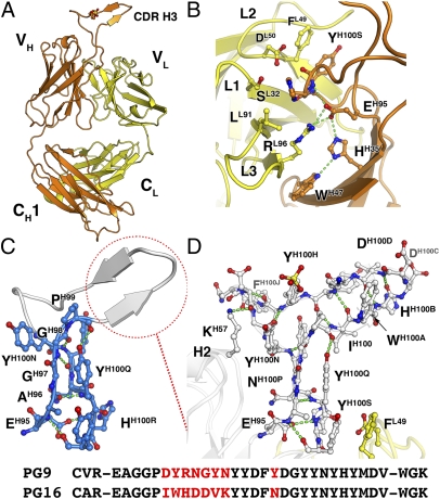
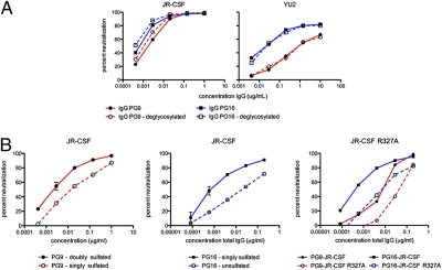
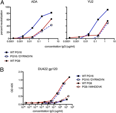
Development of an effective vaccine against HIV-1 will likely require elicitation of broad and potent neutralizing antibodies against the trimeric surface envelope glycoprotein (Env). Monoclonal antibodies (mAbs) PG9 and PG16 neutralize approximately 80% of HIV-1 isolates across all clades with extraordinary potency and target novel epitopes preferentially expressed on Env trimers. As these neutralization properties are ideal for a vaccine-elicited antibody response to HIV-1, their structural basis was investigated. The crystal structure of the antigen-binding fragment (Fab) of PG16 at 2.5 A resolution revealed its unusually long, 28-residue, complementarity determining region (CDR) H3 forms a unique, stable subdomain that towers above the antibody surface. A 7-residue "specificity loop" on the "hammerhead" subdomain was identified that, when transplanted from PG16 to PG9 and vice versa, accounted for differences in the fine specificity and neutralization of these two mAbs. The PG16 electron density maps also revealed that a CDR H3 tyrosine was sulfated, which was confirmed for both PG9 (doubly) and PG16 (singly) by mass spectral analysis. We further showed that tyrosine sulfation plays a role in binding and neutralization. An N-linked glycan modification is observed in the variable light chain, but not required for antigen recognition. Further, the crystal structure of the PG9 light chain at 3.0 A facilitated homology modeling to support the presence of these unusual features in PG9. Thus, PG9 and PG16 use unique structural features to mediate potent neutralization of HIV-1 that may be of utility in antibody engineering and for high-affinity recognition of a variety of therapeutic targets.


Conflict of interest statement

Conflict of interest statement: L.M.W, S.K.P, P.P, and D.R.B. are inventors on a patent describing the human broadly neutralizing antibodies PG9 and PG16 (United States provisional patent application numbers USSN 61/161,010; USSN 61/165,829; and USSN 61/224,739).

Figures

Fig. 1.
Fig. 1.
Overall structure of PG16 Fab illustrating the protrusion of CDR H3 from the combining site. (A) Schematic of the PG16 Fab structure with the heavy and light chains shown in orange and yellow ribbon representations, respectively. (B) Expanded view of H3 base. Heavy chain (orange) interactions with light chain (yellow) at the base of H3 are depicted. A prominent bulge at the H3 base is stabilized by interactions between GluH95 and the backbone amide of HisH100R and supported by interactions of GluH95 with ArgL96 and HisH35 and by stacking of TyrH100Q with PheL49. (C) Expanded view of H3 stalk region (marine blue). Protrusion of CDR H3 from the variable domain interface is mediated by GlyH97-GlyH98, which are sandwiched between TyrH100N and TyrH100Q to form a stalk. The polypeptide takes a sharp turn at ProH99 immediately followed by the variable region loop, which forms a short antiparallel β-sheet. (D) Full view of H3. The apex of H3 is composed of LysH100F, TyrH100G, TyrH100H, and AspH100I that display distorted β-strand geometry as they cross over the stalk region. TyrH100H is sulfated and the sulfate moiety is ordered (Fig. S3). AspH100I, PheH100J, AsnH100K, and AspH100L form a 310-helical turn. (Bottom) Alignment of the CDR H3 sequences of PG9 and PG16. The seven sequential variable residues that form the short β-sheet “specificity loop” (dashed red circle) are shown in red, as is the only other CDR H3 residue (H100K) that differs between PG9 and PG16.
Fig. 2.
Fig. 2.
Role of posttranslational modifications in neutralization of HIV-1 by PG9 and PG16. (A) Effect of enzymatic deglycosylation of PG9 and PG16 IgGs on neutralization of JR-CSF and YU2 pseudovirus. Curves shown are for control PG9 IgG (solid black circles, solid red line) and PG16 IgG (solid black squares, solid blue line) and deglycosylated PG9 IgG (open black circles, dashed red line) and PG16 IgG (open black squares, dashed blue line). Endo F treatment of PG9 and PG16 slightly increased neutralization potency against JR-CSF, but had no effect on neutralization of YU2. (B) Effect of tyrosine sulfation on neutralization of JR-CSF. Neutralization mediated by a sample of PG9 Fab enriched in the hypersulfated (PG9-S2) form (solid black circles, solid red line) was compared with a pool containing only the hyposulfated form (PG9-S) (open black circles, dashed red line). Neutralization mediated by sulfated PG16 Fab (solid black squares, solid blue line) was compared with nonsulfated PG16 Fab (open black squares, dashed blue line). For both PG9 and PG16, the more sulfated species is the more potent. Neutralization of WT JR-CSF by PG9 (solid black circles, solid red line) and PG16 IgG (solid black squares, solid blue line) was compared with neutralization of JR-CSF containing an R327A substitution by PG9 (open black circles, dashed red line) and PG16 (open black squares, dashed blue line).
Fig. 3.
Fig. 3.
Neutralization and binding data for the PG9 and PG16 CDR H3-swap variant antibodies. (A) Neutralization of PG16-hypersensitive strains ADA and YU2 by WT PG16 IgG (solid black squares, solid blue line), WT PG9 IgG (solid black circles, solid red line), and the PG16/DYRNGYN swap variant (open squares, dashed blue line). Swapping of PG9 H3 variable residues into PG16 confers a neutralization profile that is closely similar to that of PG9. (B) Recognition of monomeric DU422 gp120 by WT and H3 swap mutants, determined by ELISA. WT PG9 (solid black circles, solid red line) and the PG16/DYRNGYN swap variant (open squares, dashed blue line) display similar binding profiles with monomeric DU422 gp120. Likewise, PG16 (solid black squares, solid blue line) and the PG9/IWHDDVK swap mutant (open circles, dashed red line) show lack of appreciable binding to DU422 gp120.

Similar articles

Cited by

References

    1. Mascola JR, et al. The National Institute of Allergy and Infectious Diseases AIDS Vaccine Evaluation Group Immunization with envelope subunit vaccine products elicits neutralizing antibodies against laboratory-adapted but not primary isolates of human immunodeficiency virus type 1. J Infect Dis. 1996;173:340–348. - PubMed
    1. Flynn NM, et al. rgp120 HIV Vaccine Study Group Placebo-controlled phase 3 trial of a recombinant glycoprotein 120 vaccine to prevent HIV-1 infection. J Infect Dis. 2005;191:654–665. - PubMed
    1. Gilbert PB, et al. Correlation between immunologic responses to a recombinant glycoprotein 120 vaccine and incidence of HIV-1 infection in a phase 3 HIV-1 preventive vaccine trial. J Infect Dis. 2005;191:666–677. - PubMed
    1. Moore JP, et al. Primary isolates of human immunodeficiency virus type 1 are relatively resistant to neutralization by monoclonal antibodies to gp120, and their neutralization is not predicted by studies with monomeric gp120. J Virol. 1995;69:101–109. - PMC - PubMed
    1. Pitisuttithum P, et al. Bangkok Vaccine Evaluation Group Randomized, double-blind, placebo-controlled efficacy trial of a bivalent recombinant glycoprotein 120 HIV-1 vaccine among injection drug users in Bangkok, Thailand. J Infect Dis. 2006;194:1661–1671. - PubMed

Publication types