

Sialidase enhances recovery from spinal cord contusion injury
- PMID: 20534525
- PMCID: PMC2895144
- DOI: 10.1073/pnas.1006683107
Sialidase enhances recovery from spinal cord contusion injury
Abstract
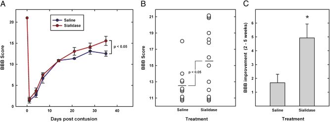
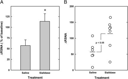
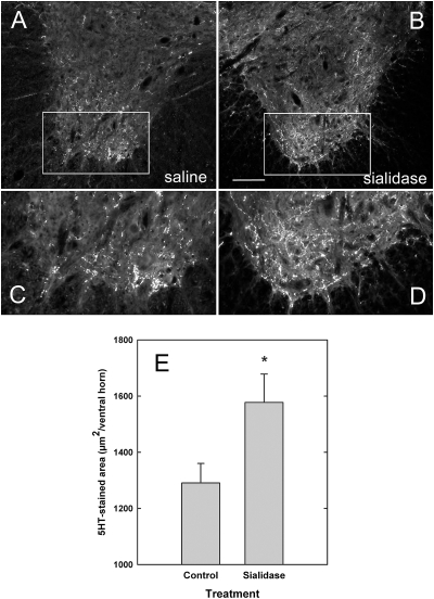
Axons fail to regenerate in the injured spinal cord, limiting motor and autonomic recovery and contributing to long-term morbidity. Endogenous inhibitors, including those on residual myelin, contribute to regeneration failure. One inhibitor, myelin-associated glycoprotein (MAG), binds to sialoglycans and other receptors on axons. MAG inhibition of axon outgrowth in some neurons is reversed by treatment with sialidase, an enzyme that hydrolyzes sialic acids and eliminates MAG-sialoglycan binding. We delivered recombinant sialidase intrathecally to rats following a spinal cord contusive injury. Sialidase (or saline solution) was infused to the injury site continuously for 2 wk and then motor behavior, autonomic physiology, and anatomic outcomes were determined 3 wk later. Sialidase treatment significantly enhanced hindlimb motor function, improved bulbospinally mediated autonomic reflexes, and increased axon sprouting. These findings validate sialoglycans as therapeutic targets and sialidase as a candidate therapy for spinal cord injury.
Conflict of interest statement
The authors declare no conflict of interest.
Figures
Similar articles
-
Sialidase, chondroitinase ABC, and combination therapy after spinal cord contusion injury.J Neurotrauma. 2013 Feb 1;30(3):181-90. doi: 10.1089/neu.2012.2353. Epub 2013 Jan 21. J Neurotrauma. 2013. PMID: 22934782 Free PMC article.
-
Ameliorative Effects of p75NTR-ED-Fc on Axonal Regeneration and Functional Recovery in Spinal Cord-Injured Rats.Mol Neurobiol. 2015 Dec;52(3):1821-1834. doi: 10.1007/s12035-014-8972-6. Epub 2014 Nov 15. Mol Neurobiol. 2015. PMID: 25394381
-
Sialidase enhances spinal axon outgrowth in vivo.Proc Natl Acad Sci U S A. 2006 Jul 18;103(29):11057-62. doi: 10.1073/pnas.0604613103. Epub 2006 Jul 17. Proc Natl Acad Sci U S A. 2006. PMID: 16847268 Free PMC article.
-
Molecular targets in spinal cord injury.J Mol Med (Berl). 2005 Sep;83(9):657-71. doi: 10.1007/s00109-005-0663-3. Epub 2005 Aug 2. J Mol Med (Berl). 2005. PMID: 16075258 Review.
-
The Nogo receptor, its ligands and axonal regeneration in the spinal cord; a review.J Neurocytol. 2002 Feb;31(2):93-120. doi: 10.1023/a:1023941421781. J Neurocytol. 2002. PMID: 12815233 Review.
Cited by
-
Expression of ligands for Siglec-8 and Siglec-9 in human airways and airway cells.J Allergy Clin Immunol. 2015 Mar;135(3):799-810.e7. doi: 10.1016/j.jaci.2015.01.004. J Allergy Clin Immunol. 2015. PMID: 25747723 Free PMC article.
-
Functional interplay between ganglioside GM1 and cross-linking galectin-1 induces axon-like neuritogenesis via integrin-based signaling and TRPC5-dependent Ca²⁺ influx.J Neurochem. 2016 Feb;136(3):550-63. doi: 10.1111/jnc.13418. Epub 2015 Dec 28. J Neurochem. 2016. PMID: 26526326 Free PMC article.
-
Neu3 sialidase-mediated ganglioside conversion is necessary for axon regeneration and is blocked in CNS axons.J Neurosci. 2014 Feb 12;34(7):2477-92. doi: 10.1523/JNEUROSCI.4432-13.2014. J Neurosci. 2014. PMID: 24523539 Free PMC article.
-
Mechanistic and Therapeutic Implications of Protein and Lipid Sialylation in Human Diseases.Int J Mol Sci. 2024 Nov 7;25(22):11962. doi: 10.3390/ijms252211962. Int J Mol Sci. 2024. PMID: 39596031 Free PMC article. Review.
-
Role of myelin-associated inhibitors in axonal repair after spinal cord injury.Exp Neurol. 2012 May;235(1):33-42. doi: 10.1016/j.expneurol.2011.05.001. Epub 2011 May 7. Exp Neurol. 2012. PMID: 21596039 Free PMC article. Review.
References
-
- Sandvig A, Berry M, Barrett LB, Butt A, Logan A. Myelin-, reactive glia-, and scar-derived CNS axon growth inhibitors: Expression, receptor signaling, and correlation with axon regeneration. Glia. 2004;46:225–251. - PubMed
-
- Cao Z, et al. Receptors for myelin inhibitors: Structures and therapeutic opportunities. Mol Cell Neurosci. 2010;43:1–14. - PubMed
-
- Fawcett JW. Overcoming inhibition in the damaged spinal cord. J Neurotrauma. 2006;23:371–383. - PubMed
Publication types
MeSH terms
Substances
Grants and funding
LinkOut - more resources
Full Text Sources
Medical
Research Materials
