. 2010 Jun 22;107(25):11495-500.
doi: 10.1073/pnas.1002099107. Epub 2010 Jun 7.

Identification of an IL-27/osteopontin axis in dendritic cells and its modulation by IFN-gamma limits IL-17-mediated autoimmune inflammation

Affiliations

Identification of an IL-27/osteopontin axis in dendritic cells and its modulation by IFN-gamma limits IL-17-mediated autoimmune inflammation

Gopal Murugaiyan et al. Proc Natl Acad Sci U S A. .

Abstract

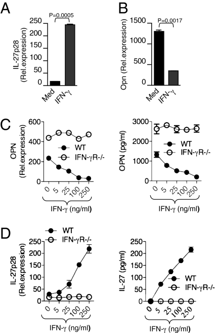
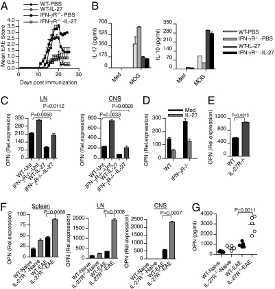
Dendritic cells (DCs) play a central role in determining the induction of T cell responses. IL-27 production by DCs favors induction of IL-10-producing regulatory T cells, whereas osteopontin (OPN) promotes pathogenic IL-17 T cell responses. The regulatory mechanisms in DCs that control these two cells types are not understood well. Here, we show that IFN-gamma induces IL-27 while inhibiting OPN expression in DCs both in vitro and in vivo and that engagement of IFN-gammaR expressed by DCs leads to suppression of IL-17 production while inducing IL-10 from T cells. DCs modified by IFN-gamma acquire IL-27-dependent regulatory function, promote IL-10-mediated T cell tolerance, and suppress autoimmune inflammation. Thus, our results identify a previously unknown pathway by which IFN-gamma limits IL-17-mediated autoimmune inflammation through differential regulation of OPN and IL-27 expression in DCs.


Conflict of interest statement

The authors declare no conflict of interest.

Figures

Fig. 1.
Fig. 1.
IFN-γ reciprocally regulates OPN and IL-27 expression in DCs. (A and B) Real-time quantitative RT-PCR analysis of mRNA encoding IL-27p28 and OPN in DCs treated with or without mouse rIFN-γ (100 ng/mL). (C and D) Dose-dependent effect of IFN-γ on IL-27 and OPN expression in DCs from WT and IFN-γR−/− mice. Shown is real-time quantitative RT-PCR analysis of mRNA encoding IL-27p28 and OPN in DCs. Data are representative of five independent experiments, and the error bars represent the mean ± SD.
Fig. 2.
Fig. 2.
IFN-γ deficiency alters OPN and IL-27 expression in DCs during EAE. (A) Real-time RT-PCR analysis of OPN in DCs isolated from spleen, LN, and CNS of naïve and EAE-bearing WT and IFN-γ−/− mice (n = 5–6 per group). (B) OPN protein expression was determined by Western blot analysis using anti-OPN Abs. (C) IL-27 expression is elevated in WT-DCs during the course of EAE. Real-time RT-PCR analysis of IL-27 in DCs isolated from spleen, LN, and CNS of naïve and EAE-bearing WT and IFN-γ−/− mice (n = 5–6 per group).
Fig. 3.
Fig. 3.
Altered OPN and IL-27 expression in DCs differentially modulate T cell production of IL-17 and IL-10. (A) Total CD4+ T cells isolated from 2D2 mice were cocultured with CD11c+ DCs isolated from naïve and MOG35–55-immunized WT and IFN-γ−/− mice. Supernatants from cultures were harvested 72 h after initiation of cultures and assayed by ELISA for IL-17 and IL-10. (B) IFN-γ treated DCs suppress IL-17 production while inducing IL-10 production from 2D2 T cells. WT and IFN-γ−/− DCs isolated from EAE mice were stimulated with IFN-γ for 12 h and were then cultured with 2D2 T cells in the presence of MOG peptide for 72 h. After 72 h, IL-17 and IL-10 production by CD4+ T cells was determined by ELISA. (C) OPN-induced IL-17 production or IL-10 inhibition from CD4+ T cells was abrogated by the addition of IL-27. CD4+ T cells were activated with anti-CD3 and anti-CD28 mAb (0.3 μg/mL) in the presence of 1 μg/mL mouse rOPN. In some culture conditions, 100 ng/mL of mouse rIL-27 was added together with 1 μg/mL mouse rOPN. Supernatants from cultures were analyzed for cytokines IL-17 and IL-10 by ELISA. Data are representative of three independent experiments, and the error bars represent the mean ± SD.
Fig. 4.
Fig. 4.
IFN-γ deficiency differentially regulates IL-17 and IL-10 expression in T cells. (A and B) Real-time RT-PCR analysis of IL-17 and IL-10 in spleen, LN, and CNS-derived T cells were isolated from WT and IFN-γ−/− mice with or without EAE (n = 8 per group). (C) LN cells derived from WT and IFN-γ−/− mice were activated in vitro with MOG35–55 (20 μg/mL) for 72 h, and cell-free supernatants from cultures were assayed for IL-17 and IL-10.
Fig. 5.
Fig. 5.
IFN-γ–modified DCs attenuate EAE in an IL-27–dependent manner. (A) DCs were left untreated (DC-Unt) or treated with IFN-γ (DC-IFN-γ) and pulsed with MOG and were then transferred into syngeneic WT mice. As indicated, some mice were coinjected with neutralizing anti-IL-27 antibody together with IFN-γ-treated DCs. Five days later, the DC primed mice were immunized with MOG and monitored for EAE. (B) LN cells obtained 18 d after immunization from mice described in (A) above, were restimulated with MOG35–55 (20 μg/mL) for 72 h. Cell-free supernatants were assayed for IL-17 and IL-10 by ELISA. (C) Clinical scores of EAE in WT and IL-27R−/− mice injected with IFN-γ–treated or –untreated DCs before MOG immunization. (D) LN cells from IFN-γ-modified DC-treated and control DC-treated WT and IL-27R−/− mice were activated in vitro with MOG35–55 (20 μg/mL) for 72 h, and cell-free culture supernatants were assayed for IL-17 and IL-10 by ELISA. (E) Clinical scores of EAE in WT mice immunized with MOG and treated with MOG-pulsed DCs modified with IFN-γ or their unmodified counterparts. Arrow indicates time of DC injection (clinical score of ≥1.5).
Fig. 6.
Fig. 6.
IL-27 treatment inhibits clinical severity of EAE in IFN-γR−/− mice. (A) IL-27 inhibits clinical severity of EAE in WT and IFN-γR−/− mice. WT and IFN-γR−/− mice (n = 6) were immunized with MOG peptide emulsified in complete freund's adjuvant. Recombinant IL-27 (0.25 μg per mouse) was administered by s.c. to immunized WT and IFN-γR−/− mice every other day from day 2 until day 18. (B) LN cells from IL-27-treated and control mice were activated in vitro with MOG35–55 (20 μg/mL) for 72 h, and cell-free culture supernatants were assayed for IL-17 and IL-10 by ELISA. (C) Real-time RT-PCR analysis of OPN in DCs isolated from LN and CNS of WT and IFN-γR−/− mice (n = 5–6 per group) treated with or without IL-27. (D) Real-time RT-PCR analysis of OPN in DCs isolated from spleen of naïve WT and IFN-γR−/− mice treated with or without rIL-27 (100 ng/mL). (E) Real-time RT-PCR analysis of OPN in DCs isolated from spleen of naïve WT and IL-27R−/− mice. (F) Real-time RT-PCR analysis of OPN in DCs isolated from spleen, LN, and CNS of naïve and EAE bearing WT and IL-27R−/− mice (n = 6 per group). (G) ELISA of serum OPN from WT and IL-27R−/− mice with or without EAE.

Similar articles

Cited by

References

    1. Kastelein RA, Hunter CA, Cua DJ. Discovery and biology of IL-23 and IL-27: Related but functionally distinct regulators of inflammation. Annu Rev Immunol. 2007;25:221–242. - PubMed
    1. Pflanz S, et al. IL-27, a heterodimeric cytokine composed of EBI3 and p28 protein, induces proliferation of naive CD4(+) T cells. Immunity. 2002;16:779–790. - PubMed
    1. Takeda A, et al. Cutting edge: Role of IL-27/WSX-1 signaling for induction of T-bet through activation of STAT1 during initial Th1 commitment. J Immunol. 2003;170:4886–4890. - PubMed
    1. Yoshimoto T, Yoshimoto T, Yasuda K, Mizuguchi J, Nakanishi K. IL-27 suppresses Th2 cell development and Th2 cytokines production from polarized Th2 cells: A novel therapeutic way for Th2–mediated allergic inflammation. J Immunol. 2007;179:4415–4423. - PubMed
    1. Wang S, Miyazaki Y, Shinozaki Y, Yoshida H. Augmentation of antigen-presenting and Th1-promoting functions of dendritic cells by WSX-1(IL-27R) deficiency. J Immunol. 2007;179:6421–6428. - PubMed

Publication types

LinkOut - more resources