. 2010 Jun 22;107(25):11620-5.
doi: 10.1073/pnas.1002912107. Epub 2010 Jun 7.

Identification of a protein required for recovery of full antenna capacity in OCP-related photoprotective mechanism in cyanobacteria

Affiliations

Identification of a protein required for recovery of full antenna capacity in OCP-related photoprotective mechanism in cyanobacteria

Clémence Boulay et al. Proc Natl Acad Sci U S A. .

Abstract

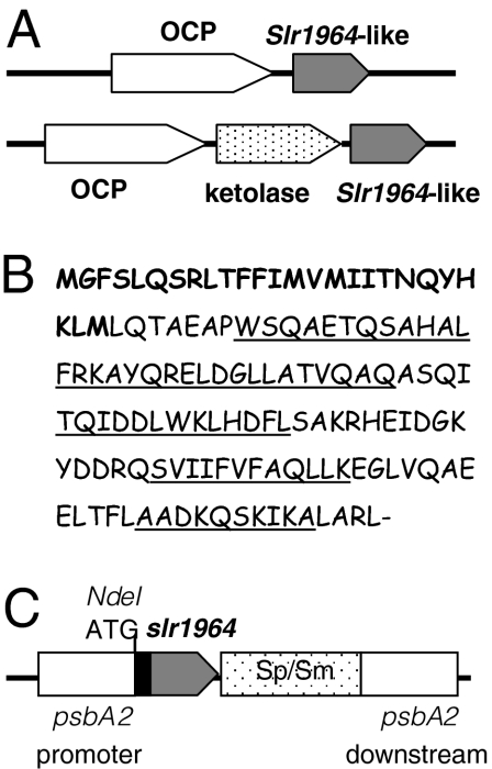
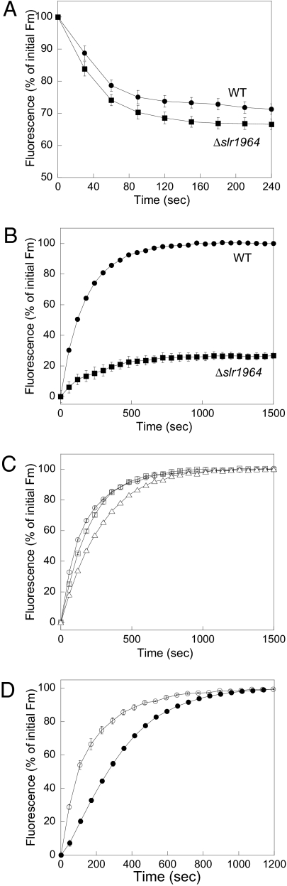
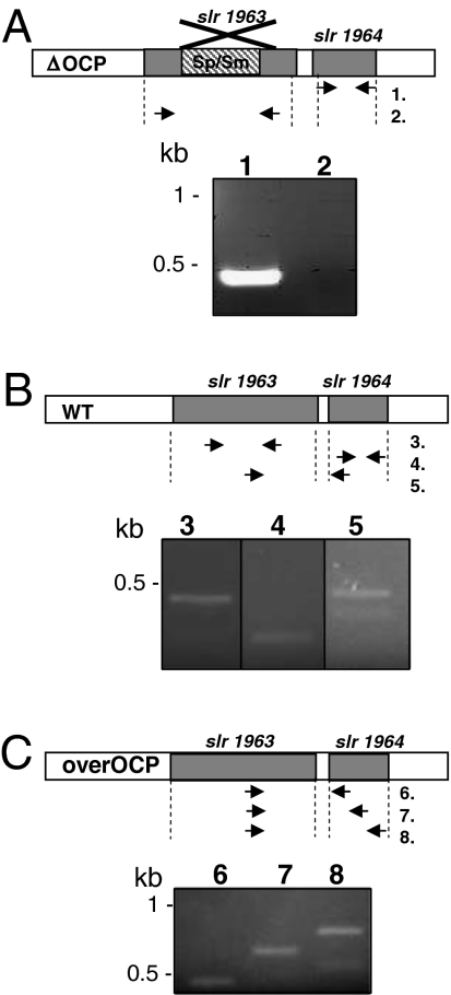
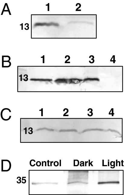
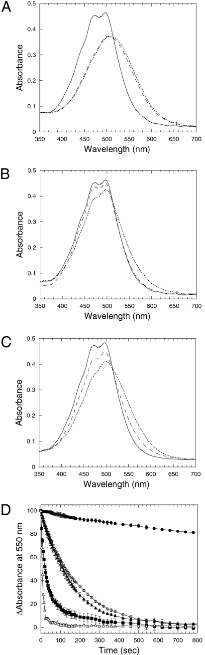
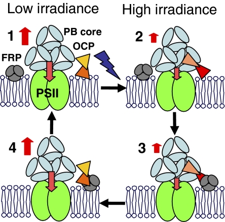
High light can be lethal for photosynthetic organisms. Similar to plants, most cyanobacteria protect themselves from high irradiance by increasing thermal dissipation of excess absorbed energy. The photoactive soluble orange carotenoid protein (OCP) is essential for the triggering of this photoprotective mechanism. Light induces structural changes in the carotenoid and the protein, leading to the formation of a red active form. Through targeted gene interruption we have now identified a protein that mediates the recovery of the full antenna capacity when irradiance decreases. In Synechocystis PCC 6803, this protein, which we called the fluorescence recovery protein (FRP), is encoded by the slr1964 gene. Homologues of this gene are present in all of the OCP-containing strains. The FRP is a 14-kDa protein, strongly attached to the membrane, which interacts with the active red form of the OCP. In vitro this interaction greatly accelerates the conversion of the red OCP form to the orange form. We propose that in vivo, FRP plays a key role in removing the red OCP from the phycobilisome and in the conversion of the free red OCP to the orange inactive form. The discovery of FRP and its characterization are essential elements in the understanding of the OCP-related photoprotective mechanism in cyanobacteria.


Conflict of interest statement

The authors declare no conflict of interest.

Figures

Fig. 1.
Fig. 1.
The slr1964 gene. (A) Gene arrangement of the slr1963-like (OCP-like) genes and slr1964-like genes in freshwater and marine cyanobacteria strains. (B) The sequence of the slr1964 Synechocystis gene. The additional N-terminal sequence existing only in Synechocystis and Microcystis is in bold. The amino acids forming the α-helices are underlined. (C) Gene arrangement in the psbA2 region of the mutant overexpressing slr1964. The slr1964 gene is under the control of the psbA2 promoter.
Fig. 2.
Fig. 2.
The role of the FRP encoded by the slr1964 gene. (A) Decrease of maximal fluorescence (Fm) during exposure of WT (circles) and Δslr1964 mutant (squares) cells to 740 μmol photons m−2 s−1 of blue-green light (400–550 nm). (BD). Increase of Fm during exposure to low blue-green light (80 μmol photons m−2 s−1) after high irradiance of cells of WT (B, circles), Δslr1964 (B, squares) overexpressing nontagged FRP (C, squares), overexpressing C-terminal His-tagged FRP (C, triangles), overexpressing N-terminal His-tagged FRP (C and D, open circles) and overexpressing OCP-FRP (D, closed circles) mutants. All curves are the averages of three independent experiments. The cells were diluted to 3 μg Chl/mL
Fig. 3.
Fig. 3.
The ocp and frp genes can be independently transcribed or cotranscribed. (A) In the ΔOCP mutant, the frp mRNA (lane 1) was present whereas the ocp mRNA was absent (lane 2); (B) In WT cells, ocp (lane 3) and frp (lane 4) mRNAs were present. An mRNA including the ocp gene and the first 30 nucleotides of the frp gene were also detectable (lane 5). (C) In the overexpressing OCP/N-terminal His-tagged FRP strain, mRNAs containing the ocp gene and at least the 30 first nucleotides of frp (lane 6) or half of the frp gene (lane 7) or the whole frp gene (lane 8) were detected. See Table S2 for primer sequences.
Fig. 4.
Fig. 4.
The location of FRP and its interaction with OCP. Immunoblot detection of FRP (A) in total proteins from the overexpressing N-terminal His-tagged FRP strain (1) and from the overexpressing OCP-FRP (N-terminal His-tagged) strain (2); (B) in total proteins (1), in the membrane-PB fraction (2), in the membrane fraction (3), and in the soluble fraction (4) isolated from the overexpressing N-terminal His-tagged FRP. (C) In isolated membranes from the overexpressing N-terminal His-tagged FRP mutant washed with Tris-HCl (1), 0.1 M Na2CO3 (2), 1 M NaCl (3), and 0.75 M NaSCN. Each lane contained 4 μg Chl (or 40 μg protein). (D) Immunoblot detection of OCP in coimmunoprecipitation experiments. The proteins attached to the Sepharose beads via the anti-FRP antibodies were eluted and separated by gel electrophoresis. OCP was then detected. The figure shows the experiment realized in darkness (dark) and under high light conditions (light, 3,000 μmol photons m−2 s−1). A control (i.e., no anti-FRP antibodies) performed under high light conditions is also shown.
Fig. 5.
Fig. 5.
FRP affects OCPr accumulation and accelerates OCPr-to-OCPo conversion. (AC) Absorbance spectra of the dark (solid line) and light forms (dashed and dotted lines) of the isolated OCP. The OCP (2.3 μM) was illuminated with white light (5,000 μmol photons m−2 s−1) in the absence (A) or presence of 2.3 μM FRP (B) or 1.15 μM FRP (C) at 18 °C (dashed line) or at 8 °C (dotted line). (D) Darkness OCPr to OCPo (2.3 μM) conversion (decrease of the absorbance at 550 nm) in the absence (circle) or in the presence of 2.3 μM FRP (square) or 1.15 μM FRP (triangle) at 8 °C (closed symbols) or at 18 °C (open symbols). Average of three independent experiments is shown.
Fig. 6.
Fig. 6.
Working model. (1) In darkness and under low irradiance the OCP is mostly orange and not (or weakly) attached to the PB. (2) Absorption of blue-green light induces changes in the carotenoid and the protein converting OCPo to OCPr. The OCPr will bind to the APC trimers of the core of the PBs via its C-terminal region and fluorescence quenching will be induced. The PB protein and chromophore interacting with the OCPr reminds to be elucidated. (3) The FRP, as a trimer, binds to the N-terminal OCPr, (4) the FRP helps the detachment of the OCPr from the PB and accelerates the OCPr-to-OCPo conversion inducing the recovery of fluorescence. Only the core of the PB is presented in the figure.

References

    1. Horton P, Ruban AV, Walters RG. Regulation of light harvesting in green plants. Annu Rev Plant Physiol Plant Mol Biol. 1996;47:655–684. - PubMed
    1. Niyogi KK. Photoprotection revisited: Genetic and molecular approaches. Annu Rev Plant Physiol Plant Mol Biol. 1999;50:333–359. - PubMed
    1. Pascal AA, et al. Molecular basis of photoprotection and control of photosynthetic light-harvesting. Nature. 2005;436:134–137. - PubMed
    1. Ruban AV, et al. Identification of a mechanism of photoprotective energy dissipation in higher plants. Nature. 2007;450:575–578. - PubMed
    1. Adir N. Elucidation of the molecular structures of components of the phycobilisome: Reconstructing a giant. Photosynth Res. 2005;85:15–32. - PubMed

Publication types