. 2010 Jun 22;107(25):11381-6.
doi: 10.1073/pnas.0906461107. Epub 2010 Jun 7.

Activity of any class IA PI3K isoform can sustain cell proliferation and survival

Affiliations

Activity of any class IA PI3K isoform can sustain cell proliferation and survival

Lazaros C Foukas et al. Proc Natl Acad Sci U S A. .

Abstract

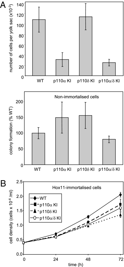
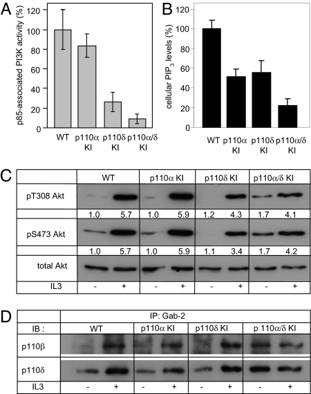
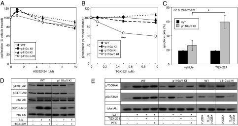
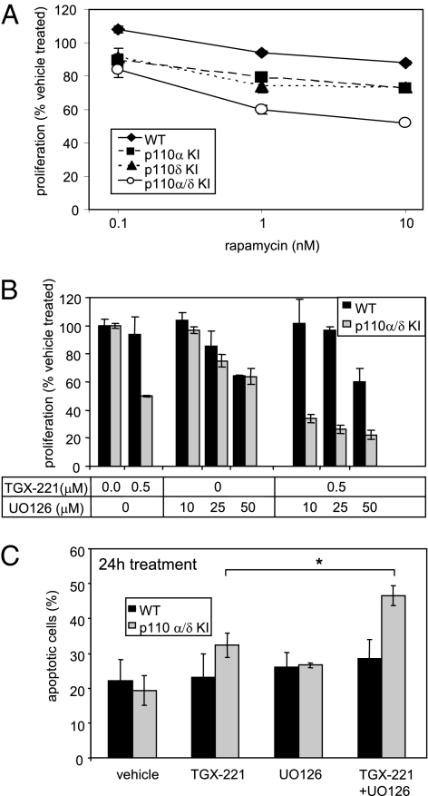
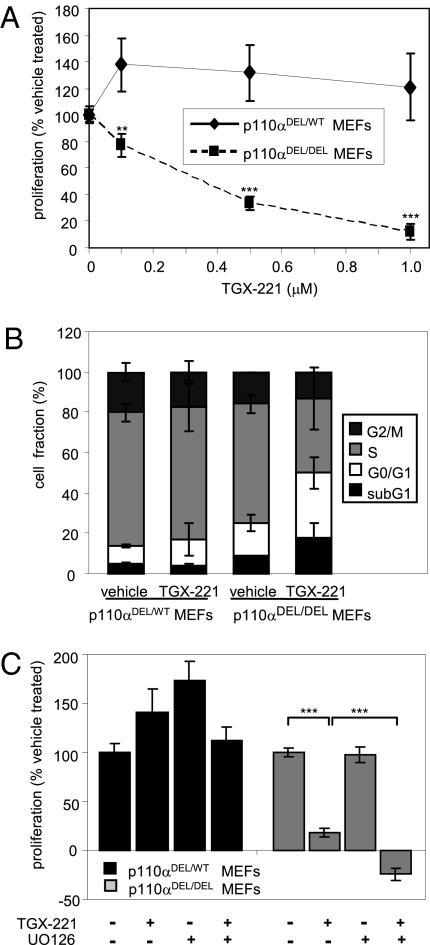
Small molecule inhibitors of PI3K for oncology mainly target the class I PI3Ks, comprising the p110alpha, beta, gamma, and delta isoforms, of which only p110alpha is mutated in cancer. To assess the roles of class I PI3K isoforms in cell proliferation and survival, we generated immortalized mouse leukocyte and fibroblast models in which class I PI3Ks were inactivated by genetic and pharmacological approaches. In IL3-dependent hemopoietic progenitor cells (which express all four class I PI3K isoforms), genetic inactivation of either p110alpha or p110delta did not affect cell proliferation or survival or sensitize to p110beta or p110gamma inactivation. Upon compound inactivation of p110alpha and p110delta, which removed >90% of p85-associated PI3K activity, remarkably, cells continued to proliferate effectively, with p110beta assuming an essential role in signaling and cell survival. Furthermore, under these conditions of diminished class I PI3K activity, input from the ERK pathway became important for cell survival. Similar observations were made in mouse embryonic fibroblasts (which mainly express p110alpha and p110beta) in which p110alpha or p110beta could sustain cell proliferation as a single isoform. Taken together, these data demonstrate that a small fraction of total class I PI3K activity is sufficient to sustain cell survival and proliferation. Persistent inhibition of selected PI3K isoforms can allow the remaining isoform(s) to couple to upstream signaling pathways in which they are not normally engaged. Such functional redundancy of class IA PI3K isoforms upon sustained PI3K inhibition has implications for the development and use of PI3K inhibitors in cancer.


Conflict of interest statement

Conflict of interest statement: Bart Vanhaesebroeck is a consultant for Intellikine (San Diego, CA).

Figures

Fig. 1.
Fig. 1.
HPC survival and proliferation upon genetic inactivation of p110α, p110δ, or both. (A) Primary cells were isolated from yolk sacs of E10.5 embryos of the designated genotypes and counted (Upper), followed by measurement of colony formation capacity in methylcellulose-based semisolid media (Lower) (equal numbers of cells were plated per condition). Pooled data from cells isolated from three embryo litters per each genotype (i.e., nine independent colony assays) are shown. (B) Immortalized Hox11-expressing HPCs of the indicated genotypes were seeded at the same density and cultured in the presence of IL3, followed by cell counting at the indicated time points. Pooled data from three independent experiments performed in triplicate are shown.
Fig. 2.
Fig. 2.
PI3K activity and signaling in immortalized HPCs upon genetic inactivation of p110α, p110δ, or both. (A) Lipid kinase activity was assayed in anti-p85 immunoprecipitates from HPC lysates. Data from one experiment performed in quadruplicate are shown. (B) Cellular PIP3 levels in IL3-stimulated HPCs. Pooled data from three independent experiments performed in duplicate are shown. The average value of p110α/δ KI HPCs treated with 1 μM TGX-221 for 16 h was subtracted as background signal. (C and D) HPCs, deprived of serum and IL3 for 3 h, were stimulated with 20 ng/mL IL3 for 5 min at 37 °C, followed by cell lysis and immunoblotting of 50 μg of total protein with the indicated antibodies to Akt (C) or by immunoprecipitation of 1.5 mg of total protein using an antibody to Gab2, followed by immunoblotting using antibodies to the indicated p110 isoforms (D). In C, relative (fold over vehicle-treated WT cells) signal intensities, indicated under each blot, were quantified by densitometry.
Fig. 3.
Fig. 3.
p110β can support survival and proliferation of p110α/δ KI cells in a largely Gβγ-independent fashion. (A and B) Dose-dependent effect of p110γ inhibitor AS252424 (A) or p110β inhibitor TGX-221 (B) on HPC proliferation assessed by MTS assay after treatment for 24 h. Pooled data from two and three independent experiments, respectively, performed in triplicate, are shown. (C) Apoptosis induced by 72-h treatment with TGX-221 (1 μM). Pooled data from four independent experiments are shown. (D) WT and p110α/δ KI HPCs cultured in the presence of 500 nM TGX-221 or vehicle for 24 h were stimulated with 20 ng/mL IL3 for 5 min at 37 °C, followed by lysis and immunoblot analysis of Akt and S6 phosphorylation using the indicated antibodies. (E) WT and p110α/δ KI HPCs cultured in serum-free RPMI medium in the presence or absence of 100 ng/mL PTX for 3 h were treated with 100 nM TGX-221 or vehicle for 30 min, followed by stimulation with 20 ng/mL IL3 for 5 min at 37 °C. Akt was analyzed by immunoblotting using the indicated antibodies. Pertussis toxin-treated WT and p110α/δ KI cells were stimulated for 5 min with the GPCR ligand SDF-1 (250 ng/mL) as a positive control for the effectiveness of PTX treatment.
Fig. 4.
Fig. 4.
mTOR and ERK pathway inhibition augments the effect of class IA PI3K inhibition on HPC survival and proliferation. (A and B) Dose-dependent effect of rapamycin (A) or TGX-221 combined with the MEK inhibitor UO126 (B) on HPC proliferation, assessed by MTS assay after 48 h. Representative experiments performed in triplicate are shown (values = mean ± SD). (C) Apoptosis in WT and p110α/δ KI HPCs upon incubation in the presence of TGX-221 (1 μM) with or without UO126 (10 μM). Data pooled from three independent experiments are shown.
Fig. 5.
Fig. 5.
Simultaneous inactivation of p110α and p110β in MEFs results in G0/G1cell cycle arrest. (A) Dose-dependent effect of the p110β inhibitor TGX-221 on proliferation of p110α-deficient (p110αDEL/DEL) MEF assessed by MTS assay after treatment for 48 h. Heterozygous p110αDEL/WT MEFs (i.e., tamoxifen-treated p110αflox/WT MEFs) were used as a control. Pooled data from three independent experiments are shown. (B) Cell cycle distribution of p110α-deficient MEFs assessed by FACS analysis of BrdU incorporation following 48-h treatment with 1 μM TGX-221. Pooled data from three independent experiments are shown. (C) Effect of combined treatment with TGX-221 (1 μM) and MEK UO126 (10 μM) on MEF proliferation, assessed by MTS assay after 48 h. Pooled data from three independent experiments are shown.

Similar articles

Cited by

References

    1. Kok K, Geering B, Vanhaesebroeck B. Regulation of phosphoinositide 3-kinase expression in health and disease. Trends Biochem Sci. 2009;34:115–127. - PubMed
    1. Shayesteh L, et al. PIK3CA is implicated as an oncogene in ovarian cancer. Nat Genet. 1999;21:99–102. - PubMed
    1. Samuels Y, et al. High frequency of mutations of the PIK3CA gene in human cancers. Science. 2004;304:554. - PubMed
    1. Vogt PK, Kang S, Elsliger MA, Gymnopoulos M. Cancer-specific mutations in phosphatidylinositol 3-kinase. Trends Biochem Sci. 2007;32:342–349. - PubMed
    1. Yuan TL, Cantley LC. PI3K pathway alterations in cancer: Variations on a theme. Oncogene. 2008;27:5497–5510. - PMC - PubMed

Publication types

Substances