. 2010 Jun 15;107(24):10914-9.
doi: 10.1073/pnas.0914067107. Epub 2010 Jun 7.

A fluorophore ligase for site-specific protein labeling inside living cells

Affiliations

A fluorophore ligase for site-specific protein labeling inside living cells

Chayasith Uttamapinant et al. Proc Natl Acad Sci U S A. .

Abstract

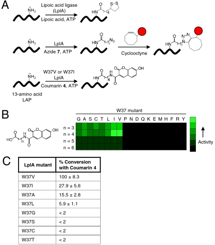
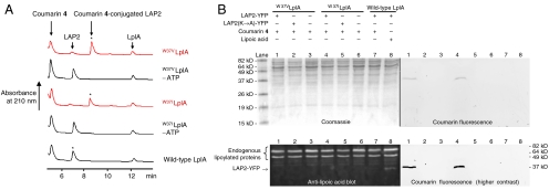
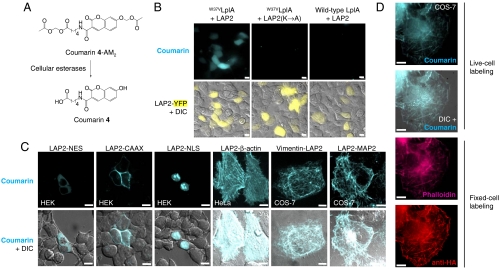
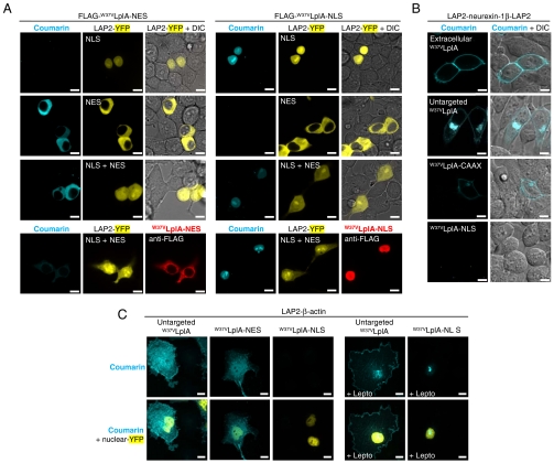
Biological microscopy would benefit from smaller alternatives to green fluorescent protein for imaging specific proteins in living cells. Here we introduce PRIME (PRobe Incorporation Mediated by Enzymes), a method for fluorescent labeling of peptide-fused recombinant proteins in living cells with high specificity. PRIME uses an engineered fluorophore ligase, which is derived from the natural Escherichia coli enzyme lipoic acid ligase (LplA). Through structure-guided mutagenesis, we created a mutant ligase capable of recognizing a 7-hydroxycoumarin substrate and catalyzing its covalent conjugation to a transposable 13-amino acid peptide called LAP (LplA Acceptor Peptide). We showed that this fluorophore ligation occurs in cells in 10 min and that it is highly specific for LAP fusion proteins over all endogenous mammalian proteins. By genetically targeting the PRIME ligase to specific subcellular compartments, we were able to selectively label spatially distinct subsets of proteins, such as the surface pool of neurexin and the nuclear pool of actin.


Conflict of interest statement

The authors declare no conflict of interest.

Figures

Fig. 1.
Fig. 1.
Engineering a coumarin ligase. (A) Natural and engineered ligation reactions catalyzed by lipoic acid ligase (LplA). The middle row shows two-step probe targeting via alkyl azide 7 ligation followed by [3 + 2] cycloaddition (12), and the bottom row shows direct fluorophore ligation by an LplA mutant. LAP = LplA Acceptor Peptide (20). The red circle represents any probe. (B) Relative activities of W37 LplA mutants with 7-hydroxycoumarin derivatives. Assays were performed under two conditions: 12 hours with LplA’s natural protein substrate E2p (15) or 40 min with LAP4.3D (20) (see SI Methods). No product was detected for the black-shaded quadrants. (C) Percent of LAP4.3D peptide converted to the coumarin (n = 4) conjugate for the eight most active W37 LplA mutants. Enzymes were used at 1 μM and reaction times were 40 min. Values are normalized to the percent conversion obtained with the best mutant, W37V, which is set to 100%. Measurements were performed in triplicate. Errors, ± 1 s.d.
Fig. 2.
Fig. 2.
In vitro characterization of coumarin ligase. (A) HPLC traces showing conversion of LAP2 peptide (20) to the coumarin 4 conjugate by W37VLplA or W37ILplA (red traces). Negative controls with wild-type LplA, or ATP omitted, are shown in black. Starred peaks were collected and analyzed by mass spectrometry (Fig. S2). (B) Coumarin labeling of mammalian cell lysate to determine sequence-specificity. Lanes 1 and 4 show coumarin 4 ligation to LAP2-YFP catalyzed by W37VLplA and W37ILplA, respectively. Negative controls are shown with wild-type LplA (lane 7), an alanine mutation in LAP2 (lanes 2 and 5), and untransfected lysate (lanes 3 and 6). The same samples were also analyzed by blotting with anti-lipoic acid antibody. Lysate in lane 8 was subjected to in vitro lipoylation with exogenous wild-type LplA.
Fig. 3.
Fig. 3.
Coumarin PRIME labeling in living mammalian cells. (A) Structure of membrane-permeant coumarin 4-AM2 probe used for intracellular labeling. Reaction catalyzed by endogenous cellular esterases produces the parent coumarin 4 structure. (B) PRIME labeling of LAP2-YFP in HEK cells. Negative controls are shown with a Lys → Ala mutation in LAP2, and with W37VLplA replaced by wild-type LplA. (C) Labeling of different LAP2 fusion proteins. NES = nuclear export sequence. CAAX = prenylation tag. NLS = nuclear localization sequence. MAP2 = microtubule-associated protein 2. (D) Comparison of methods for actin visualization. Coumarin PRIME labeling was performed on live cells expressing HA-LAP2-β-actin. After fixation, actin was detected by staining with phalloidin-Alexa Fluor 647 and anti-HA antibody. For BD, coumarin labeling was performed for 10 min with W37VLplA, followed by 1-hour washout of excess probe. All scale bars, 10 μm. Epifluorescence images are shown in B and D; confocal images are shown in C.
Fig. 4.
Fig. 4.
Compartmentalized PRIME labeling with genetically targeted coumarin ligase. (A) Compartmentalized labeling of cytosolic or nuclear LAP2-YFP. HEK cells expressing both LAP2-YFP-NES and LAP2-YFP-NLS (third row), or either construct alone (first and second rows), were labeled with coumarin using nuclear-excluded (left) or nuclear-targeted (right) ligase. On the bottom, anti-FLAG staining of fixed cells shows the expression pattern of FLAG-W37VLplA. (B) Compartmentalized labeling of the membrane protein neurexin-1β. HEK cells expressing double-LAP2 tagged neurexin-1β were labeled with W37VLplA added to the media (first row), expressed in the cytoplasm (second row), anchored to the inner plasma membrane (third row), or targeted to the nucleus (fourth row). (C) Compartmentalized labeling of actin. COS-7 cells expressing LAP2-β-actin and untargeted, nuclear-excluded, or nuclear-localized W37VLplA were labeled with coumarin for 30 min. The right panels show labeling in the presence of 2 ng/ml leptomycin B (Lepto) to inhibit nuclear export. All images in AC are confocal. Scale bars, 10 μm.

Comment in

Similar articles

Cited by

References

    1. Lin MZ, Wang L. Selective labeling of proteins with chemical probes in living cells. Physiology. 2008;23:131–141. - PubMed
    1. Los GV, et al. HatoTag: A novel protein labeling technology for cell imaging and protein analysis. ACS Chem Biol. 2008;3:373–382. - PubMed
    1. Gautier A, et al. An engineered protein tag for multiprotein labeling in living cells. Chem Biol. 2008;15:128–136. - PubMed
    1. Gallagher SS, Sable JE, Sheetz MP, Cornish VW. An in vivo covalent TMP-tag based on proximity-induced reactivity. ACS Chem Biol. 2009;4:547–556. - PMC - PubMed
    1. Moritz OL, Tam BM, Papermaster DS, Nakayama T. A functional rhodopsin-green fluorescent protein fusion protein localizes correctly in transgenic Xenopus laevis retinal rods and is expressed in a time-dependent pattern. J Biol Chem. 2001;276:28242–28251. - PubMed

Publication types

MeSH terms