. 2010 Jun 1;107(22):10026-31.
doi: 10.1073/pnas.0915137107. Epub 2010 May 13.

Ras signaling requires dynamic properties of Ets1 for phosphorylation-enhanced binding to coactivator CBP

Affiliations

Ras signaling requires dynamic properties of Ets1 for phosphorylation-enhanced binding to coactivator CBP

Mary L Nelson et al. Proc Natl Acad Sci U S A. .

Abstract

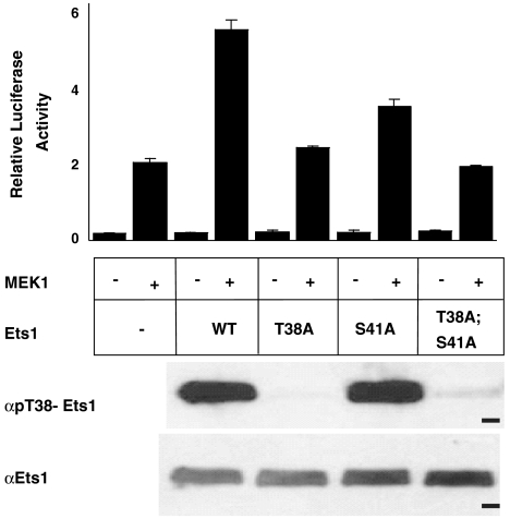
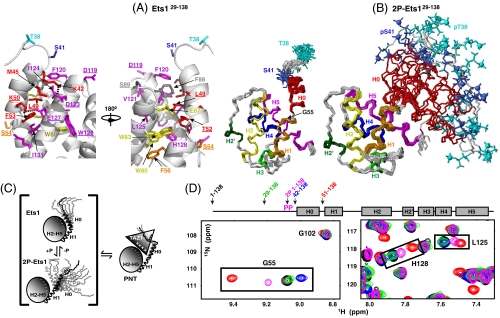
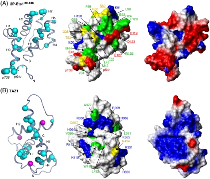
Ras/MAPK signaling is often aberrantly activated in human cancers. The downstream effectors are transcription factors, including those encoded by the ETS gene family. Using cell-based assays and biophysical measurements, we have determined the mechanism by which Ras/MAPK signaling affects the function of Ets1 via phosphorylation of Thr38 and Ser41. These ERK2 phosphoacceptors lie within the unstructured N-terminal region of Ets1, immediately adjacent to the PNT domain. NMR spectroscopic analyses demonstrated that the PNT domain is a four-helix bundle (H2-H5), resembling the SAM domain, appended with two additional helices (H0-H1). Phosphorylation shifted a conformational equilibrium, displacing the dynamic helix H0 from the core bundle. The affinity of Ets1 for the TAZ1 (or CH1) domain of the coactivator CBP was enhanced 34-fold by phosphorylation, and this binding was sensitive to ionic strength. NMR-monitored titration experiments mapped the interaction surfaces of the TAZ1 domain and Ets1, the latter encompassing both the phosphoacceptors and PNT domain. Charge complementarity of these surfaces indicate that electrostatic forces act in concert with a conformational equilibrium to mediate phosphorylation effects. We conclude that the dynamic helical elements of Ets1, appended to a conserved structural core, constitute a phospho-switch that directs Ras/MAPK signaling to downstream changes in gene expression. This detailed structural and mechanistic information will guide strategies for targeting ETS proteins in human disease.


Conflict of interest statement

The authors declare no conflict of interest.

Figures

Fig. 1.
Fig. 1.
Ras/MAPK signaling enhances Ets1 transactivation. NIH 3T3 cells were transfected with an ETS site-driven firefly luciferase reporter, an expression vector for FLAG-tagged Ets1 (wild type or the indicated mutant), and either an expression vector for constitutively active MEK1 (+) or an empty vector control (-). In addition, Renilla luciferase control vector was included to provide RLA. The bar graph (Upper) shows a representative experiment with RLA as mean and standard deviation for triplicate transfections. MEK1-dependent activation, observed in the absence of transfected Ets1, is possibly due to ERK2 effects on other elements of the transcription machinery (35). Gel panels are immunoblots showing Ets1 species from transfected cells, which were immunoprecipitated with FLAG-specific antibodies, then probed by immunoblotting with pThr38-Ets1 (Upper) or Ets1 (Lower) specific antibodies. Dash, 50 kDa marker. Additional expression controls are shown in Fig. S8G.
Fig. 2.
Fig. 2.
Superimposed structural ensembles of (A Right) Ets129-138 and (B) 2P-Ets129-138 showing the helical bundle PNT domain preceded by a flexible region containing the ERK2 phosphoacceptors Thr38 and Ser41. The α-helices (H0, residues 42–52; H1, 54–62; H2, 74–87; H3, 102–107; H4, 109–116; H5, 119–134) and a 310-helix (H2′, 95–98) are identified by rainbow coloring, and Thr38 (cyan) and Ser41 (blue) phosphates as spheres. Disordered residues 29–37 are omitted for clarity. (A Left) Expanded view of the interface between the dynamic helix H0 and helices H2 and H5 of the Ets129-138 core PNT domain, colored by helix identity. Dashes show potential salt bridges (K42-D123, K50-E127). Residues examined by mutation are underlined (see Fig. 4 and Fig. S8). (C) The phospho-switch model for the interaction of Ets1 and CBP. The PNT domain exists in a conformational equilibrium with the dynamic helix H0 in closed and open states (as well as being frayed or fully unfolded; not drawn). Phosphorylation shifts the population distribution to the open state, which is favored for TAZ1 binding via complementary electrostatic interactions. (D) This model is supported by colinear, population-weighted amide chemical shifts of residues at the helix H0/H1 junction (Gly55) and the H0 interface along H5 (Leu125 and His128) in superimposed spectra of a series of deletion fragments. The shifts of 2P-Ets11-138 (magenta) are intermediate between those of the Ets11-138 (black) and Ets129-138 (green), which are preferentially closed, and those of Ets151-138 (red), which lacks H0 and thus models fully open. The shifts of Gly55 indicate that Ets142-138 (blue) may adopt an even more closed state (see Figs. S5 and S8 for details).
Fig. 4.
Fig. 4.
Residues of Ets1 functional in transcriptional superactivation. Superactivation (RLAWT or mutant)/(RLAempty vector) is reported as mean values for three to six independent experiments ± the standard error of the mean, except mutants W126A and D123A, which had two replicates. Fig. S8 presents controls for expression levels, phosphorylation state of Thr38, and folding competence (L49R, D123R, and E127R only). Structural environments of the residues are shown in Fig. 2.
Fig. 3.
Fig. 3.
Identification of the Ets1/CBP binding interface. (Left) Residues in 2P-Ets11-138 and TAZ1 showing large 15N-HSQC intensity reductions in the presence of equimolar TAZ1 or 2P-Ets11-138, respectively (∼90% saturation; Figs. S6 and S7), are mapped on ribbon diagrams of low-energy members of the (A) 2P-Ets129-138 (representing 2P-Ets11-138) and (B) TAZ1 (PDB ID code 1u2n) structural ensembles. Increasing cyan sphere size corresponds to greater signal loss upon complex formation (intensity ratios: 0 < large sphere < 0.05, 0.05 < medium < 0.10, 0.10 < small < 0.15), and qualitatively defines the binding interface between the Ets1 and CBP fragments. Zn+2 ions, magenta balls. (Center) Surface representations of 2P-Ets129-138 and TAZ1 with residues shown as spheres now identified by physicochemical properties (red, Asp, Glu; blue, Arg, Lys, His; yellow, neutral polar; green, hydrophobic). Residues examined by mutation (Fig. 4 and Fig. S8) are underlined. (Right) Electrostatic surfaces of 2P-Ets129-138 and TAZ1, calculated with MolMol, are complementary (default parameters with “simple charge”; red, negative; blue, positive).

Similar articles

Cited by

References

    1. Tootle TL, Rebay I. Post-translational modifications influence transcription factor activity: A view from the ETS superfamily. Bioessays. 2005;27:285–298. - PubMed
    1. Li QJ, et al. MAP kinase phosphorylation-dependent activation of Elk-1 leads to activation of the co-activator p300. EMBO J. 2003;22:281–291. - PMC - PubMed
    1. Bassuk AG, Leiden JM. A direct physical association between ETS and AP-1 transcription factors in normal human T cells. Immunity. 1995;3:223–237. - PubMed
    1. Stacey KJ, Fowles LF, Colman MS, Ostrowski MC, Hume DA. Regulation of urokinase-type plasminogen activator gene transcription by macrophage colony-stimulating factor. Mol Cell Biol. 1995;15:3430–3441. - PMC - PubMed
    1. Yang BS, et al. Ras-mediated phosphorylation of a conserved threonine residue enhances the transactivation activities of c-Ets1 and c-Ets2. Mol Cell Biol. 1996;16:538–547. - PMC - PubMed

Publication types

MeSH terms