. 2010 Jun 1;107(22):10166-71.
doi: 10.1073/pnas.0909112107. Epub 2010 May 13.

SH2 domain containing leukocyte phosphoprotein of 76-kDa (SLP-76) feedback regulation of ZAP-70 microclustering

Affiliations

SH2 domain containing leukocyte phosphoprotein of 76-kDa (SLP-76) feedback regulation of ZAP-70 microclustering

Hebin Liu et al. Proc Natl Acad Sci U S A. .

Abstract

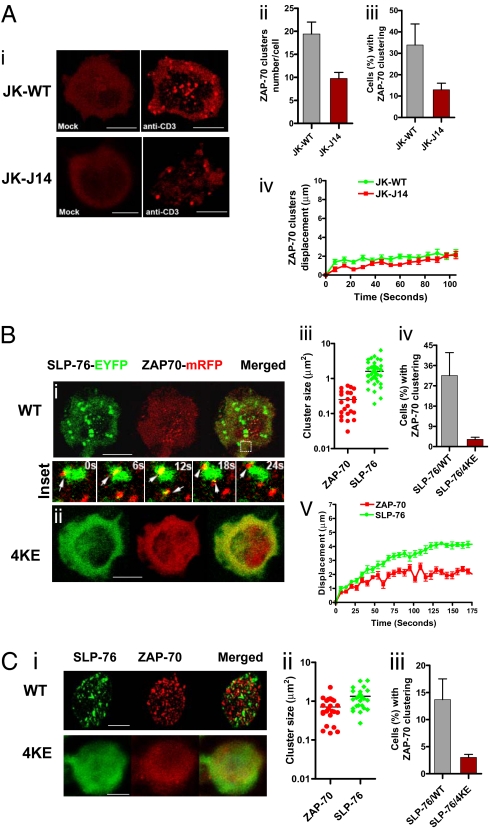
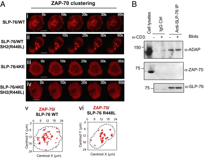
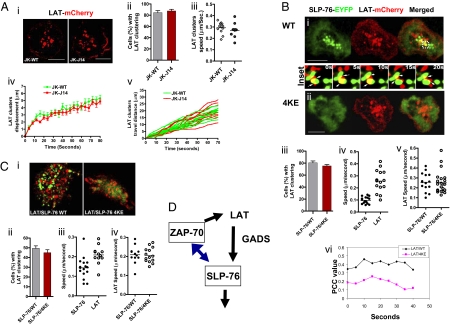
T cell receptor (TCR) signaling involves CD4/CD8-p56lck recruitment of ZAP-70 to the TCR receptor, ZAP-70 phosphorylation of LAT that is followed by LAT recruitment of the GADS-SLP-76 complex. Back regulation of ZAP-70 by SLP-76 has not been documented. In this paper, we show that anti-CD3 induced ZAP-70 cluster formation is significantly reduced in the absence of SLP-76 (i.e., J14 cells) and in the presence of a mutant of SLP-76 (4KE) in Jurkat and primary T cells. Both the number of cells with clusters and the number of clusters per cell were reduced. This effect was not mediated by SLP-76 SH2 domain binding to ZAP-70 because SLP-76 failed to precipitate ZAP-70 and an inactivating SH2 domain mutation (i.e., R448L) on SLP-76 4KE did not reverse the inhibition of ZAP-70 clustering. Mutation of R448 on WT SLP-76 still supported ZAP-70 clustering. Intriguingly, by contrast, LAT clustering occurred normally in the absence of SLP-76, or the presence of 4KE SLP-76 indicating that this transmembrane adaptor can operate independently of ZAP-70-GADS-SLP-76. Our findings reconfigure the TCR signaling pathway by showing SLP-76 back-regulation of ZAP-70, an event that could ensure that signaling components are in balance for optimal T cell activation.


Conflict of interest statement

The authors declare no conflict of interest.

Figures

Fig. 1.
Fig. 1.
SLP-76 regulates ZAP-70 microcluster formation. (A) SLP-76 deficient J14 cells show reduced levels of ZAP-70-mRFP clustering relative to SLP-76 positive WT cells. Confocal images of ZAP-70-mRFP cluster formation and distribution at the interface of transfected Jurkat WT or J14 cells and anti-CD3–coated slides (i). Jurkat WT cells expressing ZAP-70-mRFP on anti-CD3–coated slides (Upper Right) or on control anti-Ig–coated slides (Upper Left); SLP-76 deficient J14 cells expressing ZAP-70-mRFP on anti-CD3–coated slides (Lower Right) or on control anti-Ig–coated slides (Lower Left). (Scale bars, 10 μm.) Histograms showing the number of ZAP-70-mRFP clusters per cell (ii), the percentage of cells expressing ZAP-70-mRFP clusters (iii) and displacement values of ZAP-70-mRFP clusters in WT (green curve) and J14 (red curve) cells over time as determined by Volocity software (iv). Values for motility were calculated by Volocity software from the time-lapse movies. (B) Inhibition of ZAP-70-mRFP clustering by coexpression of SLP-76 4KE mutant. Confocal images of ZAP-70-mRFP (red) and SLP-76-EYFP (green) microclustering at the interface between transfected J14 cells and anti-CD3–coated coverslips. J14 cells were cotransfected with ZAP-70-mRFP and SLP-76-EYFP WT (i) or ZAP-70-mRFP and SLP-76-EYFP 4KE mutant (ii). (Insets) The transient interactions between ZAP-70-mRFP microclusters and SLP-76-EYFP WT microclusters in selected regions over 24 s. (Scale bars, 10 μm.) Histograms show cluster sizes of ZAP-70-mRFP and SLP-76-EYFP WT (iii), percentage of cells with ZAP-70 clustering in presence of SLP-76 WT or 4KE mutant expression (iv) and displacement values of ZAP-70 clusters (red curve) and SLP-76 clusters (green curve) over 175 s (v). (C) SLP-76 4KE impairs ZAP-70-mRFP cluster formation in human primary T cells. Confocal images of ZAP-70-mRFP (red) and SLP-76-EYFP (green) microclustering at the interface between transfected human primary T cells and anti-CD3–coated coverslips. Human primary T cells were cotransfected with ZAP-70-mRFP and SLP-76-EYFP WT (i, Upper) or ZAP-70-mRFP and SLP-76-EYFP 4KE mutant (i, Lower). (Scale bars, 5 μm.) Histograms show cluster size of ZAP-70-mRFP and SLP-76-EYFP (ii) and percent of cells with ZAP-70 clusters in SLP-76 and SLP-76 4KE cotransfected cells (iii).
Fig. 2.
Fig. 2.
Putative SLP-76 SH2 domain binding to ZAP-70 is detectable and cannot account for SLP-76 regulation of ZAP-70 clustering (A) Inactivation of the SH2 domain in the SLP-76 4KE mutant failed to alter its ability to disrupt ZAP-70 clustering. Time-lapse confocal images of ZAP-70 microclusters formation at the interface between transfected J14 Jurkat T cells and antigenic coverslips. ZAP-70-mRFP was coexpressed in cells with SLP-76 WT (i), SLP-76-SH2-(R448L) (ii), SLP-76–4KE (iii) or in SLP-76–4KE-SH2 (R448L) (iv). Panels are representative of at least eight cells. (Scale bars, 10 μm.) The movement of ZAP-70 individual cluster in cells over expressing SLP-76 WT (v) or SLP-76 R448L (vi) over the time course was tracked. The dotted line indicates boundary of T cell/coverslip interface. (B) SLP-76 fails to coprecipitate ZAP-70. SLP-76 was immunoprecipitated by anti-SLP-76 monoclonal antibody from Jurkat cells in the presence or absence of anti-CD3. The immunoprecipitates were subjected to SDS/PAGE, immunoblotted using anti-ADAP (Top); anti-ZAP-70 (Middle); anti-SLP-76 (Bottom).
Fig. 3.
Fig. 3.
The SLP-76 4KE mutant disrupts GADS microcluster formation. Confocal images of GADS-mCherry and SLP-76-EYFP cluster formation and distribution at the interface of transfected cells and anti-CD3–coated slides (A). Jurkat cells were cotransfected with and GADS-mCherry (red) and SLP-76-EYFP WT (green) (Upper) or with GADS-mCherry and SLP-76-EYFP 4KE mutant (Lower). (Scale bars, 10 μm.) Insets show time-lapse series of GADS and SLP-76 WT microcluster movement in selected regions imaged over 50 s. Histogram shows the speed (μm/s) traveled by individual clusters over time within cells cotransfected with SLP-76-EYFP WT and GADS-mCherry (B).
Fig. 4.
Fig. 4.
LAT clustering occurs independently of SLP-76. (A) LAT-mCherry clustering in Jurkat WT or SLP-76 deficient J14 cells. Confocal images of LAT-mCherry cluster formation and distribution at the interface of transfected cells and anti-CD3–coated slides (i) LAT-mCherry microcluster formation in Jurkat WT cells (Left) or in SLP-76 deficient J14 cells (Right). (Scale bars, 10 μm.) Histograms show the percentage of cells expressing LAT-mCherry clusters in WT (gray bar) and J14 (red bar) cells (ii) values of speed of movement of LAT clusters (iii), displacement values of LAT-mCherry clusters (iv) and distance traveled over time by individual LAT clusters (v) in WT Jurkat cells or J14 cells. Values for motility were calculated by Volocity software from the time-lapse movies. (B) LAT clustering in J14 cells cotransfected with SLP-76 WT or 4KE mutant. Confocal images of LAT-mCherry and SLP-76-EYFP cluster formation and distribution at the interface of transfected J14 cells and anti-CD3–coated slides. J14 cells were cotransfected with LAT-mCherry (red) and SLP-76-EYFP WT (green) (i) or with LAT-mCherry (red) and SLP-76-EYFP 4KE mutant (ii). Insets between i and ii show the movement and interaction of LAT-mCherry and SLP-76-EYFP clusters in selected regions over 20 s. (Scale bars, 10 μm.) Histograms show the percentage of cells with LAT clusters in Jurkat cells cotransfected with SLP-76-EYFP WT (gray bar) or SLP-76-EYFP 4KE mutant (red bar) (iii), the speed (μm/s) traveled by individual SLP-76 versus LAT clusters in cells coexpressed with LAT-mCherry and SLP-76-EYFP WT (iv), the speed of LAT clusters in cells cotransfected SLP-76-EYFP WT or 4KE mutant (v) and Pearson's correlation coefficient (PCC) values of the overlap of LAT-mCherry and SLP-76-EYFP WT or LAT-mCherry and SLP-76-EYFP 4KE over a time-course (vi). Values for motility and PCC were calculated by Volocity software from the time-lapse movies. (C) LAT clustering is not affected in primary T cells expressing SLP-76 4KE mutant. Confocal images of LAT-mCherry and SLP-76-EYFP cluster formation and distribution at the interface of transfected human primary T cells and anti-CD3–coated slides (i). Human primary T cells were cotransfected with LAT-mCherry (red) and SLP-76-EYFP WT (green) (Left) or with LAT-mCherry (red) and SLP-76-EYFP 4KE mutant (green) (Right). Histograms show the percentage of cells with LAT clusters in human primary T cells cotransfected with SLP-76-EYFP WT or 4KE mutant (ii), the speed (μm/s) traveled by individual SLP-76 versus LAT clusters in cells coexpressed with LAT-mCherry and SLP-76-EYFP WT (iii) and the speed of LAT clusters in cells cotransfected SLP-76-EYFP WT or 4KE mutant (iv). Values for motility were calculated by Volocity software from the time-lapses movies. (D) Feedback regulation of ZAP-70 clustering by SLP-76, whereas LAT clustering is independent. Phosphorylation of LAT is needed to recruit GADS and SLP-76 into the LAT signalosome. Disruption of LAT signaling can interfere with clustering of GADS and LAT. Similarly, disruption of GADS affects SLP-76 clustering. We show that ZAP-70 clustering is partially dependent on SLP-76. LAT to GADS to SLP-76 is therefore not a strictly linear pathway but involves a feedback loop where ZAP-70 and GADS clustering is dependent on SLP-76. By contrast, LAT clustering is unaffected by the loss of SLP-76 clustering.

Similar articles

Cited by

References

    1. Samelson LE. Signal transduction mediated by the T cell antigen receptor: The role of adapter proteins. Annu Rev Immunol. 2002;20:371–394. - PubMed
    1. Rudd CE. Adaptors and molecular scaffolds in immune cell signaling. Cell. 1999;96:5–8. - PubMed
    1. Jordan MS, Singer AL, Koretzky GA. Adaptors as central mediators of signal transduction in immune cells. Nat Immunol. 2003;4:110–116. - PubMed
    1. Zhang W, Sloan-Lancaster J, Kitchen J, Trible RP, Samelson LE. LAT: The ZAP-70 tyrosine kinase substrate that links T cell receptor to cellular activation. Cell. 1998;92:83–92. - PubMed
    1. Berry DM, Nash P, Liu SK, Pawson T, McGlade CJ. A high-affinity Arg-X-X-Lys SH3 binding motif confers specificity for the interaction between Gads and SLP-76 in T cell signaling. Curr Biol. 2002;12:1336–1341. - PubMed

MeSH terms

LinkOut - more resources