. 2010 Aug 13;285(33):25259-68.
doi: 10.1074/jbc.M110.145953. Epub 2010 Jun 9.

Copper homeostasis in Salmonella is atypical and copper-CueP is a major periplasmic metal complex

Affiliations

Copper homeostasis in Salmonella is atypical and copper-CueP is a major periplasmic metal complex

Deenah Osman et al. J Biol Chem. .

Abstract

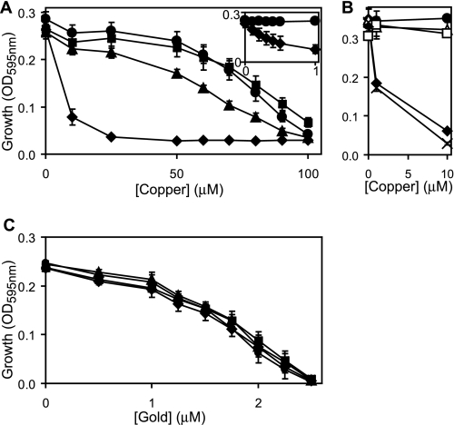
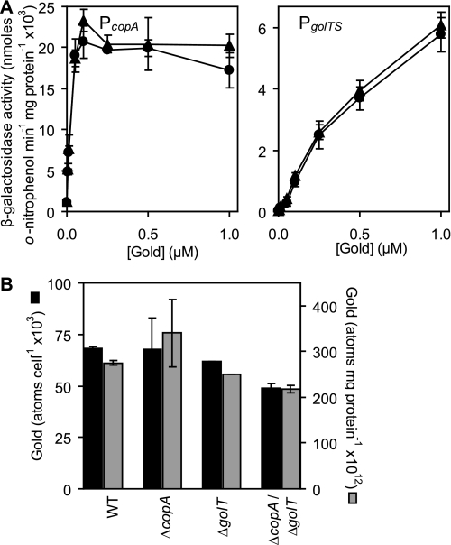
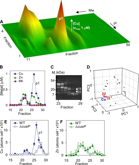
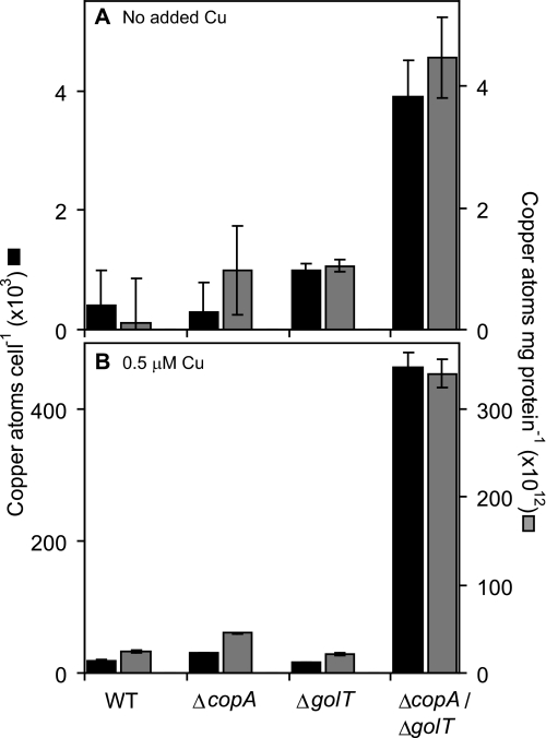
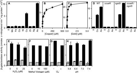
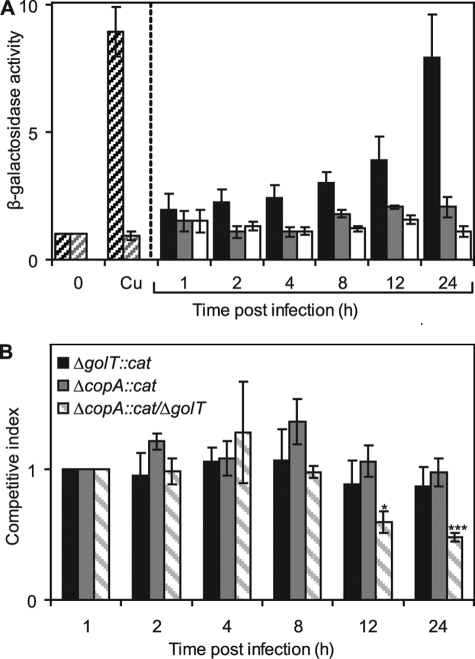
Salmonella enterica sv. typhimurium (S. enterica sv. Typhimurium) has two metal-transporting P(1)-type ATPases whose actions largely overlap with respect to growth in elevated copper. Mutants lacking both ATPases over-accumulate copper relative to wild-type or either single mutant. Such duplication of ATPases is unusual in bacterial copper tolerance. Both ATPases are under the control of MerR family metal-responsive transcriptional activators. Analyses of periplasmic copper complexes identified copper-CueP as one of the predominant metal pools. Expression of cueP was recently shown to be controlled by the same metal-responsive activator as one of the P(1)-type ATPase genes (copA), and copper-CueP is a further atypical feature of copper homeostasis in S. enterica sv. Typhimurium. Elevated copper is detected by a reporter construct driven by the promoter of copA in wild-type S. enterica sv. Typhimurium during infection of macrophages. Double mutants missing both ATPases also show reduced survival inside cultured macrophages. It is hypothesized that elevated copper within macrophages may have selected for specialized copper-resistance systems in pathogenic microorganism such as S. enterica sv. Typhimurium.


Figures

FIGURE 1.
FIGURE 1.
Both copA and golT have a role in copper export. A–C, final A595 nm measurements of wild-type S. enterica sv. Typhimurium (filled circles), ΔcopA (filled triangles), ΔgolT (filled squares), ΔcopAgolT (filled diamonds), ΔcopAgolT containing copA on plasmid pACYC184 (open triangles), ΔcopAgolT containing golT on plasmid pACYC184 (open squares) or ΔcopAgolT containing pACYC184 alone (crosses) following growth (4 h) in M9 minimal medium supplemented with increasing concentrations of copper (A and B) or gold (C). Inset, A595 nm (y axis) of wild-type S. enterica sv. Typhimurium (circles) and ΔcopAgolT (diamonds) following growth with up to 1 μm copper (x axis).
FIGURE 2.
FIGURE 2.
Cytoplasmic gold levels are unaffected by GolT. A, β-galactosidase activity was measured in wild-type S. enterica sv. Typhimurium (circles) or ΔgolT (triangles) containing PcopA or PgolTS fused to lacZ following growth in M9 minimal medium supplemented with a range of permissive gold levels. B, gold contents of wild-type S. enterica sv. Typhimurium (WT), ΔcopA, ΔgolT, and ΔcopAgolT grown in M9 minimal medium in the presence of 1 μm added gold. Metal contents are shown as atoms cell−1 (black) or atoms mg−1 cellular protein (gray).
FIGURE 3.
FIGURE 3.
The distribution of protein-bound periplasmic copper in S. enterica sv. Typhimurium. A, periplasmic extracts from wild-type S. enterica sv. Typhimurium were resolved by anion exchange (pI) into 1-ml fractions, then aliquots (0.2 ml) of 4-fold concentrated eluant resolved by size exclusion (Mr) chromatography into 0.5 ml fractions and analyzed for metals by inductively coupled plasma-mass spectrometry; the full profile for copper is shown and similar profiles were obtained with three independent extracts. B, [copper], [zinc], and [manganese] in fractions 15–31 (0.5 ml) following size exclusion of proteins eluted by 100 mm NaCl during anion exchange, which includes the major copper complex p1. C and D, the abundances of individual proteins in fractions corresponding to p1 were estimated by integrating peak areas from scanned SDS-PAGE gels of these fractions, visualized by Sypro Ruby (C), followed by principal component analysis, comparing the rise and fall of copper with the rise and fall of each protein (D). Two proteins (19 and 22) closely matching the p1 copper profile were excised from the gel and 19 was identified by mass fingerprinting as STM3650 (CueP), while 22 was not identified. E and F, the copper and zinc content of fractions 15–31 (0.5 ml) obtained following size exclusion of proteins eluted by 100 mm NaCl during anion exchange of periplasmic proteins from wild-type S. enterica sv. Typhimurium and ΔcueP shown as atoms cell−1, data points represent the mean (±S.E.) for three independent experiments.
FIGURE 4.
FIGURE 4.
Mutants lacking CopA and GolT accumulate copper. Copper contents of wild-type S. enterica sv. Typhimurium (WT), ΔcopA, ΔgolT, and ΔcopAgolT grown in M9 minimal medium in the absence (A) or presence (B) of 0.5 μm added copper. Metal contents are shown as atoms cell−1 (black) or atoms mg−1 cellular protein (gray). Note the different axes scales for A and B.
FIGURE 5.
FIGURE 5.
Metal-responsive expression from PcopA is unaffected by pH or reactive oxygen species. A–C, β-galactosidase activity measured in S. enterica sv. Typhimurium containing PcopA fused to lacZ following growth (150 min) in LB medium with no metal supplement or 1 mm Mn(II), 1 mm Fe(III), or maximum permissive concentrations of Co(II) (0.1 mm), Ni(II) (0.5 mm), Cu(II) (0.5 mm), Zn(II) (1 mm), Ag(I) (2 μm), or Au(III) (5 μm) (A), or up to inhibitory concentrations of Cu(II) (B) or Au(III) (C). D, expression from PcopA in S. enterica sv. Typhimurium (WT), ΔcueR, ΔgolS or ΔgolScueR following growth in LB medium with no metal supplement or maximum permissive concentrations of Cu(II) or Au(III). E–H, expression from PcopA in S. enterica sv. Typhimurium following growth (150 min) in LB medium with no metal supplement (gray) or with added copper (25 μm) to achieve ∼half-maximal copper-induced expression (black), with or without hydrogen peroxide (E) or the intracellular superoxide generator methyl viologen (F), or prior to washing and exposure (1 h) to fresh medium with or without 0.1 units ml−1 of the extracellular superoxide generator xanthine oxidase (G) or at different pH (H). Expression from a control promoter, PpolA (40), was unaffected by any of the conditions tested (data not shown).
FIGURE 6.
FIGURE 6.
Copper export is important for intracellular survival of S. enterica sv. Typhimurium. A, macrophages (RAW264.7) were infected with wild-type S. enterica sv. Typhimurium (black) or the ΔgolScueR derivative (gray) containing PcopA fused to lacZ following growth in M9 minimal medium and β-galactosidase activity was measured in bacteria isolated at indicated time points postinfection. β-Galactosidase activity was also measured in wild-type S. enterica sv. Typhimurium containing PcopA-lacZ maintained in M9 minimal medium (extracellular) for the duration of the infection (non-filled). Expression levels in bacteria used for the infection, grown with no added Cu(II) or grown in parallel with 25 μm Cu(II) are also shown (wild-type, dark diagonal shading; ΔgolScueR, light diagonal shading). B, competitive infections were performed with wild-type S. enterica sv. Typhimurium and either ΔcopA::cat (gray), ΔgolT::cat (black), or ΔcopA::cat/ΔgolT (diagonal shading), with the competitive index defined as the colony-forming unit ratio of mutant and wild-type strains recovered at the indicated time points postinfection, divided by their ratio in the input (intracellular bacteria after the initial treatment with gentamicin, 1-h postinfection). In each case, bacterial strains were mixed at a ratio of 1:1 and used at a final MOI of 10:1 (bacteria:macrophage). Data points represent the mean (±S.E.) for at least four independent experiments, each performed in triplicate (***, p < 0.000005; *, p < 0.005 by Student's t test).

References

    1. Coburn B., Grassl G. A., Finlay B. B. (2007) Immunol. Cell Biol. 85, 112–118 - PubMed
    1. Majowicz S. E., Musto J., Scallan E., Angulo F. J., Kirk M., O'Brien S. J., Jones T. F., Fazil A., Hoekstra R. M. (2010) Clin. Infect. Dis. 50, 882–889 - PubMed
    1. Fields P. I., Swanson R. V., Haidaris C. G., Heffron F. (1986) Proc. Natl. Acad. Sci. U.S.A. 83, 5189–5193 - PMC - PubMed
    1. Mastroeni P., Vazquez-Torres A., Fang F. C., Xu Y., Khan S., Hormaeche C. E., Dougan G. (2000) J. Exp. Med. 192, 237–248 - PMC - PubMed
    1. Vazquez-Torres A., Jones-Carson J., Mastroeni P., Ischiropoulos H., Fang F. C. (2000) J. Exp. Med. 192, 227–236 - PMC - PubMed

Publication types