. 2010 Jul;137(14):2365-74.
doi: 10.1242/dev.047605. Epub 2010 Jun 9.

Myocardin-related transcription factors regulate the Cdk5/Pctaire1 kinase cascade to control neurite outgrowth, neuronal migration and brain development

Affiliations

Myocardin-related transcription factors regulate the Cdk5/Pctaire1 kinase cascade to control neurite outgrowth, neuronal migration and brain development

Mayssa H Mokalled et al. Development. 2010 Jul.

Abstract

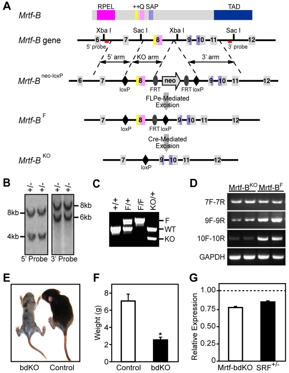
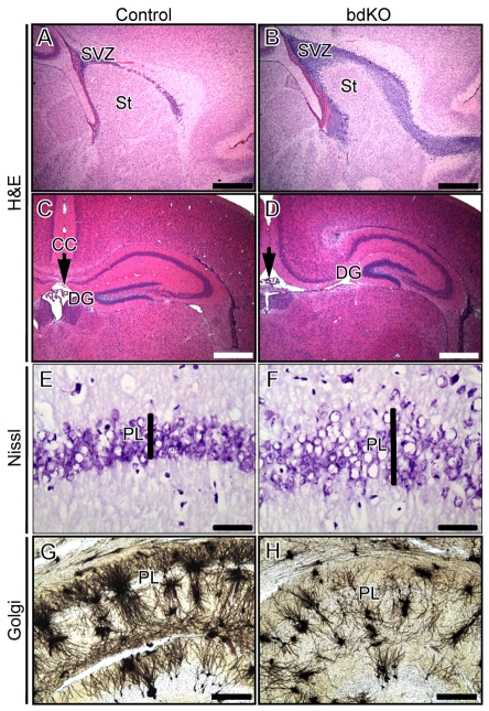
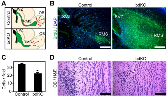
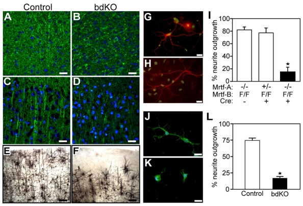
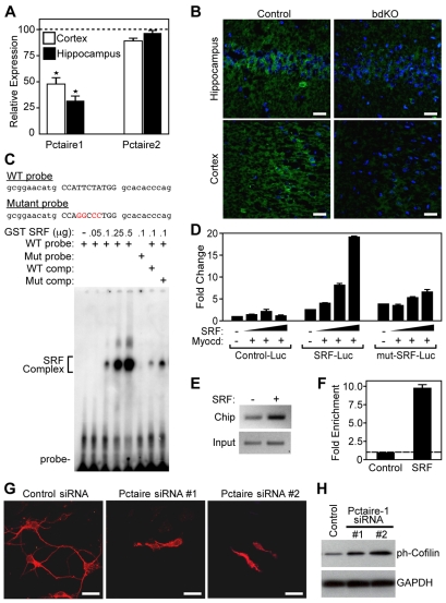
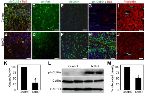
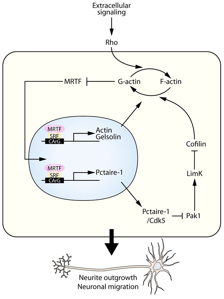
Numerous motile cell functions depend on signaling from the cytoskeleton to the nucleus. Myocardin-related transcription factors (MRTFs) translocate to the nucleus in response to actin polymerization and cooperate with serum response factor (Srf) to regulate the expression of genes encoding actin and other components of the cytoskeleton. Here, we show that MRTF-A (Mkl1) and MRTF-B (Mkl2) redundantly control neuronal migration and neurite outgrowth during mouse brain development. Conditional deletion of the genes encoding these Srf coactivators disrupts the formation of multiple brain structures, reflecting a failure in neuronal actin polymerization and cytoskeletal assembly. These abnormalities were accompanied by dysregulation of the actin-severing protein gelsolin and Pctaire1 (Cdk16) kinase, which cooperates with Cdk5 to initiate a kinase cascade that governs cytoskeletal rearrangements essential for neuron migration and neurite outgrowth. Thus, the MRTF/Srf partnership interlinks two key signaling pathways that control actin treadmilling and neuronal maturation, thereby fulfilling a regulatory loop that couples cytoskeletal dynamics to nuclear gene transcription during brain development.


Figures

Fig. 1.
Fig. 1.
Generation of MRTF brain double-knockout (bdKO) mice. (A) Strategy to generate a conditional Mrtfb allele. The protein, corresponding exonic structure, and targeted alleles are shown. The targeted Mrtfbneo-loxP allele was generated by homologous recombination in ES cells. To generate the MrtfbF allele, the neomycin resistance cassette, flanked by FRT sites, was removed by crossing to FLPe transgenic mice. The MrtfbF allele includes two loxP sites inserted into introns 7 and 8. The MrtfbKO allele was generated using Cre-mediated excision, which results in one loxP site in place of exon 8. (B) Southern blot analysis confirming targeting of ES cells. The corresponding wild-type (8 kb) and targeted bands (4 kb and 6 kb) are indicated for the 5′ and 3′ probes. (C) Genotyping of MrtfbF and MrtfbKO mice by genomic PCR. Primer set includes three primers, with two flanking the 5′ loxP site and the third downstream of the 3′ loxP site. (D) MRTF knockout in MRTF bdKO brains compared with control brains was assayed by RT-PCR using primers located within exons 7, 9 and 10 of Mrtfb. (E) MRTF bdKO mouse (left) and control Mrtfa−/+;MrtfbF/F;GFAP-Cre mouse (right) at P14. Note that one copy of Mrtfa or Mrtfb is sufficient for normal development. (F) Average weight of control and bdKO mice at P14. The MRTF bdKO mice are significantly smaller than their control littermates. (G) Expression of Srf transcripts in MRTF bdKO brains. Shown is a quantitative RT-PCR transcript analysis from the cortical regions of MRTF bdKO and Srf+/− mice. Relative expression was normalized to the corresponding transcript levels in control brains.
Fig. 2.
Fig. 2.
Neuroanatomical defects in MRTF bdKO mice. (A,B) Hematoxylin and Eosin (H&E)-stained brain sections of control (A) and MRTF bdKO (B) brains at P16. Note the reduced striatum (St) and the broad subventricular zone (SVZ). (C,D) H&E-stained coronal brain sections of control (C) and bdKO (D) mice reveal a missing corpus callosum (CC) (arrowheads) and a deformed hippocampus with deformed dentate gyrus (DG). (E-H) Defective hippocampal anatomy of MRTF bdKO mice. Nissl staining reveals a loosely defined pyramidal cell layer (PL) of the hippocampi of MRTF bdKO mice (F) compared with their control littermates (E). Golgi staining reveals defects in the arrangement of the pyramidal neurons and diminished neuronal projections within the hippocampi in the MRTF bdKO brains (H) compared with control brain sections (G). Scale bars: 500 μm in A-D; 100 μm in E-H.
Fig. 3.
Fig. 3.
Defects in neuronal cell migration from the SVZ. (A) Migration of neuronal precursors along the rostral migratory stream (RMS). Schematic illustrates the normal migration of cells born at the SVZ along the RMS to reach the olfactory bulbs (OB), where they differentiate into interneurons. This process is disrupted in bdKO mice, in which cells accumulate at the SVZ. (B) BrdU labeling shows impaired migration of neuronal cells from the SVZ along the RMS. Sagittal brain sections stained with anti-BrdU antibody reveal the accumulation of BrdU-positive cells in the SVZ of MRTF bdKO brains as compared with normal migration in the control sections. (C) Cell counts for neurons at the olfactory bulb in MRTF bdKO mice. Counts of the number of cells per field (40 fields total from four sections) reveal a significant difference between the number of neurons in the olfactory bulb of bdKO and control mice (*P<0.0001). (D) H&E-stained brain sections at the level of the olfactory bulb show a decrease in the number of interneurons in the bdKO mice as compared with their control littermates. Scale bars: 200 μm.
Fig. 4.
Fig. 4.
Decreased neuronal projections in MRTF bdKO brains. (A-D) Reduced MAP2 staining in the striatum (A,B) and cortical (C,D) regions of MRTF bdKO mice at P16. Shown are anti-MAP2 antibody-stained sections of control and MRTF bdKO mice. (E,F) Defective anatomy of the cerebellar cortex of MRTF bdKO mice. Golgi staining reveals defects in the arrangement of cortical neurons and decreased neuronal projections in the MRTF bdKO brains (F) compared with control brain sections (E). (G-I) Hippocampal neurons derived from Mrtfa−/+;MrtfbF/F or Mrtfa−/−;MrtfbF/F mouse pups at P0-P3 were infected with either GFP-Cre-expressing (H) or control GFP-expressing (G) adenovirus. Shown are the GFP and anti-MAP2 antibody stainings from 2-week neuron cultures. (I) The percentage of GFP-positive cells that show neurite outgrowth. A significant decrease in the percentage of neurite outgrowth is seen in Mrtfa−/−;MrtfbF/F Cre-infected neurons compared with Mrtfa−/−;MrtfbF/F GFP-infected or Mrtfa−/+;MrtfbF/F Cre-infected control neurons. Forty-five fields were counted from three different experiments (*P<0.03). (J-L) Neurite outgrowth in hippocampal neurons derived from MRTF bdKO or control Mrtfa−/+;MrtfbF/F mouse pups. Shown is anti-MAP2 antibody staining from 1-week neuron cultures. (L) Significant decrease in the percentage of neurite outgrowth in the bdKO culture compared with the control culture. Thirty fields were counted from three different experiments (*P<0.001). Scale bars: 50 μm in A-D; 100 μm in E,F; 10 μm in G,H,J,K.
Fig. 5.
Fig. 5.
Identification of Pctaire1 as a novel MRTF/Srf target gene. (A) Decreased expression of Pctaire1 transcripts in MRTF bdKO brains. Quantitative RT-PCR transcript analysis from hippocampal and cortical regions of MRTF bdKO brains. Relative expression was normalized to the corresponding transcript levels in control brains. *P<0.05. (B) Diminished levels of Pctaire1 protein in MRTF bdKO brains. Anti-Pctaire1 antibody staining revealed decreased levels of the protein in the hippocampal and cortical regions of MRTF bdKO mice compared with control mice. (C) Gel mobility shift assays were performed with a labeled Pctaire1 probe and purified GST-Srf or control GST proteins. Increased binding of GST-Srf to the Pctaire1 CArG sequence was detected with increased amounts of recombinant Srf. The use of a labeled mutant probe or an unlabeled probe blocks Srf binding. (D) Responsiveness of Pctaire1 expression to MRTF/Srf signaling. Increased amounts of the Srf were able to activate the Pctaire1-luciferase reporter constructs containing the predicted Srf binding site but not the empty luciferase constructs. Mutation of the predicted CArG box blocks this activation. (E,F) Enrichment of Srf at its predicted binding site within the PCTAIRE1 gene. ChIP assay were performed on chromatin prepared from human 293T cells transfected with either a flag-tagged Srf construct or a control empty construct. The precipitated genomic DNA was analyzed by RT-PCR (E) and by qPCR (F) using primers for the human PCTAIRE1 gene. Shown also is the PCR amplification performed prior to immunoprecipitation as the input control (E). (G) Defective neurite outgrowth in Pctaire1 knockdown neurons. Anti-MAP2 antibody staining from 1-week neuron cultures shows disruption of neurite outgrowth in the Pctaire1 siRNA-transfected but not in control siRNA-transfected neurons. (H) Increased cofilin phosphorylation in Pctaire1 knockdown neurons. Western blot analysis of phospho-cofilin and total cofilin in control and Pctaire1 siRNA-transfected neurons. Gapdh was used as a loading control. Scale bars: 50 μm in B; 5 μm in G.
Fig. 6.
Fig. 6.
Regulation of actin dynamics by the MRTF/Srf pathway. (A-H) Dysregulation of phosphorylated (ph) Cdk5, LimK and cofilin in MRTF bdKO brains. Anti-ph-Cdk5 (A,B), anti-ph-Pak (C,D), anti-ph-LimK (E,F), and anti-ph-cofilin (G,H) staining revealed decreased phosphorylation of Cdk5 and increased phosphorylation of LimK and cofilin in the brains of MRTF bdKO mice (B,D,F,H) as compared with control mice (A,C,E,G). (I,J) Decreased actin polymerization in the MRTF bdKO brain. Phalloidin staining revealed decreased levels of F-actin in the hippocampal regions of MRTF bdKO mice (J) compared with control mice (I). Scale bars: 50 μm. (K) Diminished kinase activity of Cdk5 upon MRTF deletion. Kinase assays show a ~75% decrease in the kinase activity of Cdk5 immunoprecipitated from the brains of MRTF bdKO mice compared with control mice. *P<0.05. (L) Western blot analysis of phospho-cofilin and total cofilin in the cerebellar cortex of control and MRTF bdKO brain samples. Gapdh was used as a loading control. (M) Quantification of F-actin levels in MRTF bdKO mice. Shown is the percentage integrated density (pixel density per area) of the phalloidin-stained MRTF bdKO cortical sections normalized to control sections. *P<0.05.
Fig. 7.
Fig. 7.
Model for the regulation of neuronal migration and neurite outgrowth by the MRTF/Srf pathway. MRTFs are essential mediators of Srf signaling in the mouse brain and key regulators of the dynamics of the actin cytoskeleton. Together with Srf, MRTFs regulate the expression of β-actin and other actin-binding proteins, such as gelsolin. In addition, activation of MRTF/Srf signaling regulates the expression of the Pctaire1 kinase, which is involved in the regulation of neurite outgrowth. Under normal conditions, the MRTF/Srf pathway regulates the activity of Cdk5, which phosphorylates and inactivates Pak1. Decreased Pak1 activity results in diminished LimK activity towards phosphorylation of cofilin, resulting in increased actin severing. Genetic ablation of MRTFs in the mouse brain disrupts actin dynamics by disrupting the expression and/or activity of actin and the actin-severing proteins gelsolin and cofilin, resulting in defective neuronal migration and neurite outgrowth.

Similar articles

Cited by

References

    1. Ahlemeyer B., Baumgart-Vogt E. (2005). Optimized protocols for the simultaneous preparation of primary neuronal cultures of the neocortex, hippocampus and cerebellum from individual newborn (P0.5) C57Bl/6J mice. J. Neurosci. Methods 149, 110-120 - PubMed
    1. Alberti S., Krause S. M., Kretz O., Philippar U., Lemberger T., Casanova E., Wiebel F. F., Schwarz H., Frotscher M., Schutz G., et al. (2005). Neuronal migration in the murine rostral migratory stream requires serum response factor. Proc. Natl. Acad. Sci. USA 102, 6148-6153 - PMC - PubMed
    1. Arber S., Barbayannis F. A., Hanser H., Schneider C., Stanyon C. A., Bernard O., Caroni P. (1998). Regulation of actin dynamics through phosphorylation of cofilin by LIM-kinase. Nature 393, 805-809 - PubMed
    1. Arsenian S., Weinhold B., Oelgeschlager M., Ruther U., Nordheim A. (1998). Serum response factor is essential for mesoderm formation during mouse embryogenesis. EMBO J. 17, 6289-6299 - PMC - PubMed
    1. Ayala R., Shu T., Tsai L. H. (2007). Trekking across the brain: the journey of neuronal migration. Cell 128, 29-43 - PubMed

Publication types

MeSH terms