Cholecystokinin is up-regulated in obese mouse islets and expands beta-cell mass by increasing beta-cell survival
- PMID: 20534724
- PMCID: PMC2940525
- DOI: 10.1210/en.2010-0233
Cholecystokinin is up-regulated in obese mouse islets and expands beta-cell mass by increasing beta-cell survival
Abstract
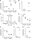
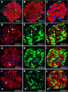
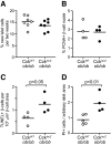
An absolute or functional deficit in beta-cell mass is a key factor in the pathogenesis of diabetes. We model obesity-driven beta-cell mass expansion by studying the diabetes-resistant C57BL/6-Leptin(ob/ob) mouse. We previously reported that cholecystokinin (Cck) was the most up-regulated gene in obese pancreatic islets. We now show that islet cholecystokinin (CCK) is up-regulated 500-fold by obesity and expressed in both alpha- and beta-cells. We bred a null Cck allele into the C57BL/6-Leptin(ob/ob) background and investigated beta-cell mass and metabolic parameters of Cck-deficient obese mice. Loss of CCK resulted in decreased islet size and reduced beta-cell mass through increased beta-cell death. CCK deficiency and decreased beta-cell mass exacerbated fasting hyperglycemia and reduced hyperinsulinemia. We further investigated whether CCK can directly affect beta-cell death in cell culture and isolated islets. CCK was able to directly reduce cytokine- and endoplasmic reticulum stress-induced cell death. In summary, CCK is up-regulated by islet cells during obesity and functions as a paracrine or autocrine factor to increase beta-cell survival and expand beta-cell mass to compensate for obesity-induced insulin resistance.
Figures


Similar articles
-
Overexpression of pre-pro-cholecystokinin stimulates beta-cell proliferation in mouse and human islets with retention of islet function.Mol Endocrinol. 2008 Dec;22(12):2716-28. doi: 10.1210/me.2008-0255. Epub 2008 Oct 9. Mol Endocrinol. 2008. Retraction in: Mol Endocrinol. 2010 Feb;24(2):472. doi: 10.1210/mend.24.2.9999. PMID: 18845673 Free PMC article. Retracted.
-
Cholecystokinin expression in the β-cell leads to increased β-cell area in aged mice and protects from streptozotocin-induced diabetes and apoptosis.Am J Physiol Endocrinol Metab. 2015 Nov 15;309(10):E819-28. doi: 10.1152/ajpendo.00159.2015. Epub 2015 Sep 22. Am J Physiol Endocrinol Metab. 2015. PMID: 26394663 Free PMC article.
-
Glucagon-Like Peptide-1 Regulates Cholecystokinin Production in β-Cells to Protect From Apoptosis.Mol Endocrinol. 2015 Jul;29(7):978-87. doi: 10.1210/me.2015-1030. Epub 2015 May 18. Mol Endocrinol. 2015. PMID: 25984632 Free PMC article.
-
beta-cell function in obese-hyperglycemic mice [ob/ob Mice].Adv Exp Med Biol. 2010;654:463-77. doi: 10.1007/978-90-481-3271-3_20. Adv Exp Med Biol. 2010. PMID: 20217510 Review.
-
The physiology of obese-hyperglycemic mice [ob/ob mice].ScientificWorldJournal. 2007 May 29;7:666-85. doi: 10.1100/tsw.2007.117. ScientificWorldJournal. 2007. PMID: 17619751 Free PMC article. Review.
Cited by
-
Association of Gut Hormones and Microbiota with Vascular Dysfunction in Obesity.Nutrients. 2021 Feb 13;13(2):613. doi: 10.3390/nu13020613. Nutrients. 2021. PMID: 33668627 Free PMC article. Review.
-
Gastrin, Cholecystokinin, Signaling, and Biological Activities in Cellular Processes.Front Endocrinol (Lausanne). 2020 Mar 6;11:112. doi: 10.3389/fendo.2020.00112. eCollection 2020. Front Endocrinol (Lausanne). 2020. PMID: 32210918 Free PMC article. Review.
-
Cholecystokinin receptor antagonist halts progression of pancreatic cancer precursor lesions and fibrosis in mice.Pancreas. 2014 Oct;43(7):1050-9. doi: 10.1097/MPA.0000000000000194. Pancreas. 2014. PMID: 25058882 Free PMC article.
-
Beneficial effects of the novel cholecystokinin agonist (pGlu-Gln)-CCK-8 in mouse models of obesity/diabetes.Diabetologia. 2012 Oct;55(10):2747-2758. doi: 10.1007/s00125-012-2654-6. Epub 2012 Jul 20. Diabetologia. 2012. PMID: 22814764
-
Vaccination with Polyclonal Antibody Stimulator (PAS) Prevents Pancreatic Carcinogenesis in the KRAS Mouse Model.Cancer Prev Res (Phila). 2021 Oct;14(10):933-944. doi: 10.1158/1940-6207.CAPR-20-0650. Epub 2021 Aug 24. Cancer Prev Res (Phila). 2021. PMID: 34429319 Free PMC article.
References
-
- Butler AE, Janson J, Bonner-Weir S, Ritzel R, Rizza RA, Butler PC 2003 β-Cell deficit and increased β-cell apoptosis in humans with type 2 diabetes. Diabetes 52:102–110 - PubMed
-
- Klöppel G, Löhr M, Habich K, Oberholzer M, Heitz PU 1985 Islet pathology and the pathogenesis of type 1 and type 2 diabetes mellitus revisited. Surv Synth Pathol Res 4:110–125 - PubMed
-
- Ritzel RA, Butler AE, Rizza RA, Veldhuis JD, Butler PC 2006 Relationship between β-cell mass and fasting blood glucose concentration in humans. Diabetes Care 29:717–718 - PubMed
-
- Keller MP, Choi Y, Wang P, Davis DB, Rabaglia ME, Oler AT, Stapleton DS, Argmann C, Schueler KL, Edwards S, Steinberg HA, Chaibub Neto E, Kleinhanz R, Turner S, Hellerstein MK, Schadt EE, Yandell BS, Kendziorski C, Attie AD 2008 A gene expression network model of type 2 diabetes links cell cycle regulation in islets with diabetes susceptibility. Genome Res 18:706–716 - PMC - PubMed
-
- Bock T, Pakkenberg B, Buschard K 2003 Increased islet volume but unchanged islet number in ob/ob mice. Diabetes 52:1716–1722 - PubMed
Publication types
MeSH terms
Substances
Grants and funding
LinkOut - more resources
Full Text Sources
Medical
Molecular Biology Databases
Miscellaneous

