. 2010 Jul 15;16(14):3684-95.
doi: 10.1158/1078-0432.CCR-09-2384. Epub 2010 Jun 9.

Development of an orthotopic model of invasive pancreatic cancer in an immunocompetent murine host

Affiliations

Development of an orthotopic model of invasive pancreatic cancer in an immunocompetent murine host

William W Tseng et al. Clin Cancer Res. .

Abstract

Purpose: The most common preclinical models of pancreatic adenocarcinoma utilize human cells or tissues that are xenografted into immunodeficient hosts. Several immunocompetent, genetically engineered mouse models of pancreatic cancer exist; however, tumor latency and disease progression in these models are highly variable. We sought to develop an immunocompetent, orthotopic mouse model of pancreatic cancer with rapid and predictable growth kinetics.

Experimental design: Cell lines with epithelial morphology were derived from liver metastases obtained from Kras(G12D/+);LSL-Trp53(R172H/+);Pdx-1-Cre mice. Tumor cells were implanted in the pancreas of immunocompetent, histocompatible B6/129 mice, and the mice were monitored for disease progression. Relevant tissues were harvested for histologic, genomic, and immunophenotypic analysis.

Results: All mice developed pancreatic tumors by two weeks. Invasive disease and liver metastases were noted by six to eight weeks. Histologic examination of tumors showed cytokeratin-19-positive adenocarcinoma with regions of desmoplasia. Genomic analysis revealed broad chromosomal changes along with focal gains and losses. Pancreatic tumors were infiltrated with dendritic cells, myeloid-derived suppressor cells, macrophages, and T lymphocytes. Survival was decreased in RAG(-/-) mice, which are deficient in T cells, suggesting that an adaptive immune response alters the course of disease in wild-type mice.

Conclusions: We have developed a rapid, predictable orthotopic model of pancreatic adenocarcinoma in immunocompetent mice that mimics human pancreatic cancer with regard to genetic mutations, histologic appearance, and pattern of disease progression. This model highlights both the complexity and relevance of the immune response to invasive pancreatic cancer and may be useful for the preclinical evaluation of new therapeutic agents.


Figures

Figure 1
Figure 1
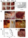
Early and late disease and in our orthotopic model. At 2 weeks, LM-P tumor uptake is noted with progressive growth at 4 weeks. Comparison of the two techniques for implantation (see Methods) reveals no difference in tumor appearance (A) or total tumor volume (B) at these early time points. By 6 to 8 weeks, mice develop extensive local growth into adjacent organs (dotted line = tumor invading into stomach) and liver metastases (open arrows) (C). In some mice, clinically relevant disease manifestations, such as biliary obstruction (arrowheads = dilated common bile duct) producing jaundice (swab tip of bilious peritoneal fluid), and gastric outlet obstruction (* = massively dilated stomach) are seen (D). In comparison to LM-P, orthotopic implantation of PDA cells results in more aggressive disease progression with decreased survival (for PDA1-1, E, *p<0.05) and development of carcinomatosis (for both PDA1-1 and PDA3-5, F).
Figure 2
Figure 2
Histology of pancreatic tumors and metastases. LM-P tumors form glands and regions of desmoplasia (A, arrows). Immunohistochemistry demonstrates that pancreatic tumors stain for cytokeratin-19, a ductal epithelial marker (B, CK19 in brown, hematoxylin counterstain in blue, original magnification, 200 X). Areas of perineural invasion (C, arrowheads = nerve, dotted line = tumor invasion) and peri-pancreatic lymph node infiltration (D) are also noted. Liver (E) and lung metastases (F) confirm adenocarcinoma. PDA pancreatic tumors show similar histology to LM-P but are predominantly poorly-(1-1, G) or well-differentiated (3-5, H) (H&E, original magnification, 100 X for A, and D-F; 200 X for G, H; 400 X for B, C; N = nerve; T = tumor, GC = germinal center)
Figure 3
Figure 3
Comparative genomic hybridization. Shown are genomic profiles of copy number alteration for LM-P cells (below, in green), a subcutaneous tumor (middle, in blue), and an orthotopic tumor (above, in red). Tumor/normal CGH log2 ratios (moving average 0.2 megabases) are plotted by chromosome position; gains and losses appear as peaks and valleys, respectively. Selected sites of gain and loss (see text) are indicated.
Figure 4
Figure 4
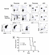
LM-P tumors are infiltrated with distinct mononuclear cell types. Three expanded populations are seen in pancreatic tumors and, to a lesser extent, in draining lymph nodes, based on surface marker expression: 1) CD11c-high, CD11b-negative 2) CD11c-high, CD11b-positive and 3) CD11c-negative/low, CD11b-positve cells (A). The two CD11c-high populations (1 and 2) express MHC Class II, and a small proportion of the cells also express CD86. The CD11c-negative/low CD11b-positive cells (3) are comprised of Gr-1+ myeloid-derived suppressor cells and Gr-1− tumor-associated macrophages. The T cell response in pancreatic tumors and draining lymph nodes is shifted toward CD4+ cells (B). Tumor-infiltrating CD4+ T cells (+) include a distinct population of CD25+ FoxP3+ regulatory T cells. Numerical values represent CD4:CD8 ratios. Isotype controls (for CD86, FoxP3) are shown in gray. Lymphocyte-deficient RAG-/- pancreatic tumor-bearing mice have decreased survival compared to fully immunocompetent mice (*p<0.05); however, both ultimately succumb to disease (C).
Figure 5
Figure 5
Treatment of LM-P tumors with gemcitabine and LY364947, a TGF-β receptor-1 kinase inhibitor (TGF-βR1 KI). In vitro, MTT assays demonstrate direct cytotoxicity of gemcitabine but not LY364947 against LM-P cells (A). In vivo, gemcitabine treatment results in improved survival, an effect slightly enhanced by addition of LY364947 (B, Gem = gemcitabine; *p<0.05 for Gem vs. PBS control **p<0.05 for Gem + TGF-βR1 KI vs. Gem alone).

Similar articles

Cited by

References

    1. Jemal A, Siegel R, Ward E, et al. Cancer statistics, 2008. CA Cancer J Clin. 2008;58:71–96. - PubMed
    1. Izeradjene K, Hingorani SR. Targets, trials, and travails in pancreas cancer. J Natl Compr Canc Netw. 2007;5:1042–53. - PubMed
    1. Hotz HG, Reber HA, Hotz B, et al. An orthotopic nude mouse model for evaluating pathophysiology and therapy of pancreatic cancer. Pancreas. 2003;26:e89–98. - PubMed
    1. Schwarz RE, McCarty TM, Peralta EA, Diamond DJ, Ellenhorn JD. An orthotopic in vivo model of human pancreatic cancer. Surgery. 1999;126:562–7. - PubMed
    1. Talmadge JE, Donkor M, Scholar E. Inflammatory cell infiltration of tumors: Jekyll or Hyde. Cancer Metastasis Rev. 2007;26:373–400. - PubMed

Publication types