Case Reports
. 2010 Sep;95(9):E58-63.
doi: 10.1210/jc.2010-0488. Epub 2010 Jun 9.

An autosomal recessive syndrome of joint contractures, muscular atrophy, microcytic anemia, and panniculitis-associated lipodystrophy

Affiliations
Case Reports

An autosomal recessive syndrome of joint contractures, muscular atrophy, microcytic anemia, and panniculitis-associated lipodystrophy

Abhimanyu Garg et al. J Clin Endocrinol Metab. 2010 Sep.

Abstract

Context: Genetic lipodystrophies are rare disorders characterized by partial or complete loss of adipose tissue and predisposition to insulin resistance and its complications such as diabetes mellitus, hypertriglyceridemia, hepatic steatosis, acanthosis nigricans, and polycystic ovarian syndrome.

Objective: The objective of the study was to report a novel autosomal recessive lipodystrophy syndrome.

Results: We report the detailed phenotype of two males and one female patient, 26-34 yr old, belonging to two pedigrees with an autosomal recessive syndrome presenting with childhood-onset lipodystrophy, muscle atrophy, severe joint contractures, erythematous skin lesions, and microcytic anemia. Other variable clinical features include hypergammaglobulinemia, hepatosplenomegaly, generalized seizures, and basal ganglia calcification. None of the patients had diabetes mellitus or acanthosis nigricans. Two had mild hypertriglyceridemia and all had low levels of high-density lipoprotein cholesterol. Skin biopsy of an erythematous nodular skin lesion from one of the patients revealed evidence of panniculitis. The lipodystrophy initially affected the upper body but later became generalized involving abdomen and lower extremities as well.

Conclusions: We conclude that these patients represent a novel autoinflammatory syndrome resulting in joint contractures, muscle atrophy, microcytic anemia, and panniculitis-induced lipodystrophy. The molecular genetic basis of this disorder remains to be elucidated.


Figures
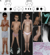
Figure 1
Figure 1
Pedigrees and clinical features of our patients with JMP syndrome. A, Pedigrees of the patients. Circles denote females and squares denote males. Arrows indicate probands. Filled symbols indicate affected and unfilled symbols indicate unaffected subjects. A diagonal line across a symbol indicates a deceased subject. A small filled circle indicates a miscarriage. B, Anterior view of patient JMP 100.3 showing marked loss of sc fat from the face, neck, chest, and upper extremities. The loss of sc fat is less evident from the abdomen and lower extremities. There is also loss of muscle mass from the upper extremities. Contractures of the upper extremities with flexion contractures at the elbows and wrists and contractures of the hands are seen. The patient has mild gynecomastia. He has no loss of scalp hair. C, JMP 200.3 showing similar features as seen in JMP100.3. In addition, there is marked loss of sc fat from the abdomen and lower extremities. The lower extremities also show marked loss of muscle mass. D, JMP 200.4 shows more marked loss of sc fat from the face, neck, chest, and upper extremities than from the abdomen, hips, and lower extremities, which were spared. The breasts are atrophic and the neck and chest showed many discrete, small, erythematous nodular skin lesions. She had mild contractures of the hand joints. E, View of the hand from patient JMP 200.3 showing flexion contractures at the elbow and wrist. The metacarpophalangeal joints were hyperextended, and the proximal and distal interphalangeal joints showed flexion contractures. F, The foot of patient JMP 200.3 showing hallux valgus deformity and flexion contractures of the rest of the toes. He also had severe inversion at the ankle joint. G, Lateral radiograph of the right hand and the forearm of patient JMP 100.3 showing flexion deformity of the wrist with poor visualization of the carpal bones. Extension deformity of the metacarpophalangeal joints and flexion deformity of proximal interphalangeal joints is seen. H, Anteroposterior radiograph of the foot of patient JMP 100.3 showing juxtaarticular osteopenia. Heads of metatarsals were unusually large with thin shafts. There was hallux valgus deformity. No acroosteolysis was seen. I, Hematoxylin and eosin stain of the skin and sc tissue biopsy specimen from JMP 200.4 showing lymphocytic infiltrate permeating deep dermal collagen and sc adipose tissue, with a perivascular component (lower left part of field) consistent with panniculitis. No vasculitis or fat necrosis is seen (×200).
Figure 2
Figure 2
MRI scans of patient JMP 200.3 in the right panels compared with a control subject (32 yr old healthy Mexican-American male with a body mass index of 21.7 kg/m2) in the left panels. A, Axial MRI of the head through the orbits shows reduction of sc fat but preservation of retroorbital fat. B, Axial MRI of the head through the nose shows reduction of sc fat from the posterior occipital region and from the malar region. C, Axial MRI of the neck just below the chin shows near total loss of sc fat from the neck. D, Sagittal MRI of the head, neck, and thorax through the orbits shows marked reduction in sc fat from the head, neck, and thorax. E, Coronal MRI of the shoulder and arm shows reduced sc fat and muscle mass. F, Coronal MRI through the abdomen and pelvis at the level of the kidneys shows marked loss sc fat from the abdomen and thighs with preservation of retroperitoneal fat. G, Axial MRI through the thorax shows marked loss of sc fat. H, Axial MRI through the abdomen at the level of the kidneys reveals marked loss of sc fat with preservation of perirenal and ip fat. I, Axial MRI through the pelvis at the level of the femoral head shows marked loss of sc fat, especially from the posterior region. Bone marrow fat and the mechanical fat around the hip joint are well preserved. Axial MRIs through the arm (J) and forearm (K) show marked loss of sc fat and reduced muscle mass. Axial MRI of the thigh (L) shows marked loss of sc fat from the posterior and lateral regions but preservation of sc fat from the anterior-medial region and of the calf (M) shows loss of sc fat from the lateral region extending to the anterior and posterior regions with preservation of sc fat in the anterior and medial region. The muscle mass in the thigh appears well preserved, but in the calf it is slightly reduced.

References

    1. Garg A 2004 Acquired and inherited lipodystrophies. N Engl J Med 350:1220–1234 - PubMed
    1. Garg A, Agarwal AK 2009 Lipodystrophies: disorders of adipose tissue biology. Biochim Biophys Acta 1791:507–513 - PMC - PubMed
    1. Tanaka M, Miyatani N, Yamada S, Miyashita K, Toyoshima I, Sakuma K, Tanaka K, Yuasa T, Miyatake T, Tsubaki T 1993 Hereditary lipo-muscular atrophy with joint contracture, skin eruptions and hyper-γ-globulinemia: a new syndrome. Intern Med 32:42–45 - PubMed
    1. Yamada S, Toyoshima I, Mori S, Tsubaki T 1984 [Sibling cases with lipodystrophic skin change, muscular atrophy, recurrent skin eruptions, and deformities and contractures of the joints. A possible new clinical entity]. Rinsho Shinkeigaku 24:703–710 - PubMed
    1. Oyanagi K, Sasaki K, Ohama E, Ikuta F, Kawakami A, Miyatani N, Miyatake T, Yamada S 1987 An autopsy case of a syndrome with muscular atrophy, decreased subcutaneous fat, skin eruption and hyper γ-globulinemia: peculiar vascular changes and muscle fiber degeneration. Acta Neuropathol 73:313–319 - PubMed

Publication types

MeSH terms