. 2010 Aug 1;21(15):2770-9.
doi: 10.1091/mbc.E09-12-1029. Epub 2010 Jun 9.

The retinoblastoma protein modulates Tbx2 functional specificity

Affiliations

The retinoblastoma protein modulates Tbx2 functional specificity

Keith W Vance et al. Mol Biol Cell. .

Abstract

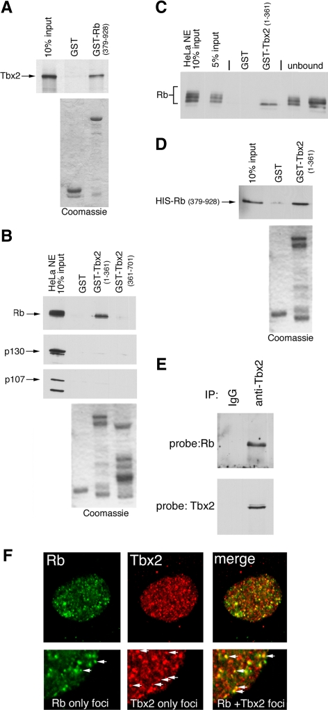
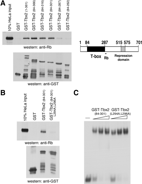
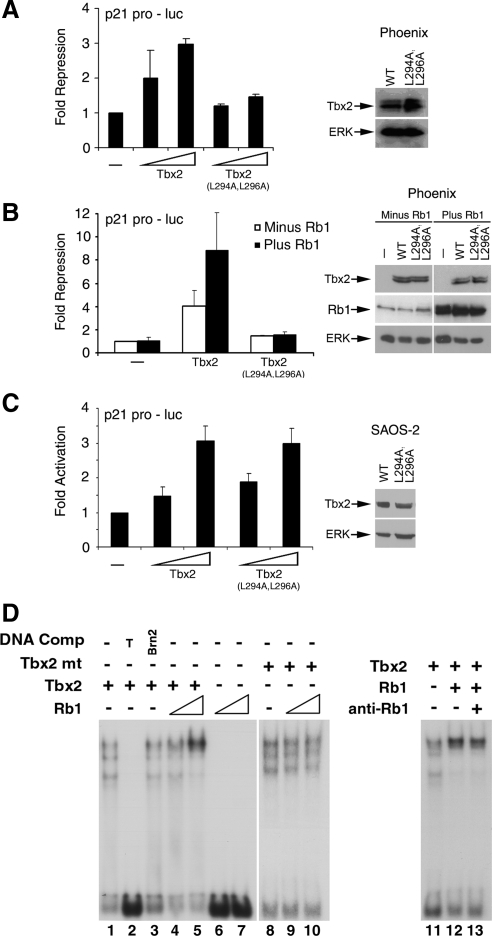
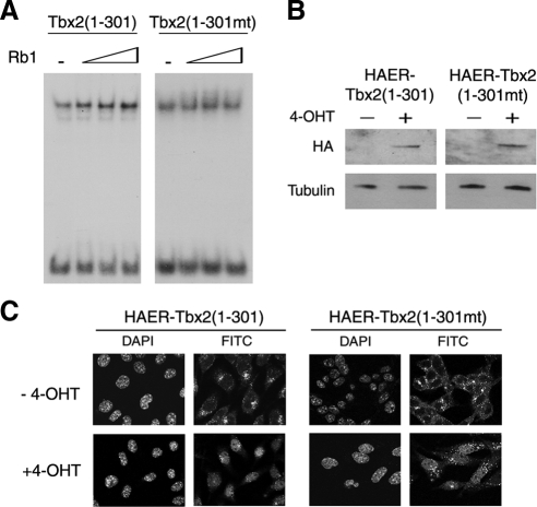
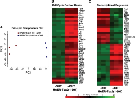
Tbx2 is a member of a large family of transcription factors defined by homology to the T-box DNA-binding domain. Tbx2 plays a key role in embryonic development, and in cancer through its capacity to suppress senescence and promote invasiveness. Despite its importance, little is known of how Tbx2 is regulated or how it achieves target gene specificity. Here we show that Tbx2 specifically associates with active hypophosphorylated retinoblastoma protein (Rb1), a known regulator of many transcription factors involved in cell cycle progression and cellular differentiation, but not with the Rb1-related proteins p107 or p130. The interaction with Rb1 maps to a domain immediately carboxy-terminal to the T-box and enhances Tbx2 DNA binding and transcriptional repression. Microarray analysis of melanoma cells expressing inducible dominant-negative Tbx2, comprising the T-box and either an intact or mutated Rb1 interaction domain, shows that Tbx2 regulates the expression of many genes involved in cell cycle control and that a mutation which disrupts the Rb1-Tbx2 interaction also affects Tbx2 target gene selectivity. Taken together, the data show that Rb1 is an important determinant of Tbx2 functional specificity.


Figures

Figure 1.
Figure 1.
Tbx2 directly interacts with hypophosphorylated Rb1. The indicated GST fusion proteins were immobilized on glutathione beads and incubated with either 35S-labeled IVT Tbx2 (A), HeLa nuclear extract (B and C), or purified HisRb(379-928) protein (D). After extensive washing, bound proteins were separated by SDS-PAGE and visualized either by autoradiography (A) or Western blotting (B–D). Tbx2 associates with Rb1 in B16 melanoma cells (E). Lysates from B16 cells were prepared and immunoprecipitated using either anti-Tbx2 or an unrelated control mAb. Proteins in the complex were identified by Western blotting using anti-Rb1 (top) or anti-Tbx2 antibody (bottom). (F) Immunofluorescence staining of B16 cells using anti-Tbx2 and anti-Rb1 antibodies after salt extraction of soluble proteins.
Figure 2.
Figure 2.
Mapping the Rb1 interaction domain in Tbx2. (A) Deletion of Tbx2 amino acids 301-287 greatly reduces binding to Rb1. (B) Identification of a Tbx2 mutant protein that fails to bind Rb1. The indicated GST-Tbx2 deletion mutants immobilized on glutathione beads were incubated with HeLa nuclear extract in a pulldown assay. Proteins were detected by Western blotting using anti-Rb1 and anti-GST antibodies. Tbx2 L294AL296A mutant protein is able to bind DNA (C). Equal amounts of purified GST-Tbx2(84-301) and GST-Tbx2(L294A,L296A) fusion proteins were incubated with radiolabeled T-element as a probe in a band-shift assay.
Figure 3.
Figure 3.
Tbx2 protein that does not bind Rb1 has an impaired transcriptional repression function (A). Phoenix cells were transfected with 50 ng p21CIP1 promoter luciferase reporter and either 25 ng or 50 ng of wild-type Tbx2 or Tbx2 L29AL296A expression vectors. Cotransfection of Rb1 enhances Tbx2-mediated transcriptional repression (B). Phoenix cells were cotransfected with 50 ng p21CIP1 promoter reporter construct along with the indicated combinations of either Tbx2 (50 ng), Tbx2mt (50 ng), and/or Rb1 (100 ng) expression vectors. Tbx2 activates transcription in Rb1 negative cells (C). SAOS-2 cells were transfected with 50 ng p21CIP1 promoter luciferase reporter and either 25 ng or 50 ng of wild-type Tbx2 or Tbx2 L29AL296A expression vectors. For these reporter experiments cells were assayed for luciferase and β-galactosidase activity 48 h after transfection. pCMV-B-gal (25 ng) was used as a transfection control and empty pCMV vector was added to make the total amount of transfected DNA equal in each case. Western blotting using anti-Tbx2, ant-Rb1, and anti-ERK antibodies was used to determine the levels of the transfected proteins. Rb1 increases the ability of Tbx2 to bind DNA (D). The indicated combinations of purified GST-Tbx2, GST-Tbx2(L294A,L296A), and His-Rb(379-928) proteins were incubated in a band-shift assay using radiolabeled T-element as a probe. Excess unlabeled T-element and Brn-2 oligos were used to assess the specificity of binding. For the supershift experiment purified proteins were incubated with 1 μl anti-Rb1 rabbit polyclonal antibody before addition of the probe.
Figure 4.
Figure 4.
Generation and characterization of B16 cell lines expressing ER-Tbx2(1-301) and ER-Tbx2(1-301mt) proteins. (A) Rb1 enhances the DNA-binding activity of a Tbx2(1-301) but not Tbx2(1-301mt) protein. Bandshift assay in which suboptimal levels of purified GST-Tbx2(1-301) and GST-Tbx2(1-301mt) fusion proteins were incubated with a consensus T-element probe and increasing concentrations of purified His-Rb(379-928). Western blotting (B) using anti-HA or anti-tubulin antibodies and immunofluorescence staining (C) with an anti-HA antibody showing the levels and subcellular localization of ER-Tbx2(1-301) and ER-Tbx2(1-301mt) proteins grown in the absence or presence of 4-OHT for 24 h.
Figure 5.
Figure 5.
Rb1 modulates Tbx2(1-301) target gene specificity. (A) PCA analysis reveals large transcriptomic differences between ER-Tbx2(1-301)– and ER-Tbx2(1-301mt)–induced cells. (B and C) Heat map displaying gene expression changes of cell cycle (GO:0007049) and transcription factor (GO:0006350) genes after ER-Tbx2(1-301) induction. Green-to-red shift indicates repressed and red-to-green shift marks up-regulated genes. Some targets are present in both categories. Genes indicated in bold are differentially expressed between ER-Tbx2(1-301)– and ER-Tbx2(1-301mt)–induced cells.

Similar articles

Cited by

References

    1. Abrahams A., Mowla S., Parker M. I., Goding C. R., Prince S. UV-mediated regulation of the anti-senescence factor Tbx2. J. Biol. Chem. 2008;283:2223–2230. - PubMed
    1. Aksan I., Goding C. R. Targeting the microphthalmia basic helix-loop-helix-leucine zipper transcription factor to a subset of E-box elements in vitro and in vivo. Mol. Cell. Biol. 1998;18:6930–6938. - PMC - PubMed
    1. Amellem O., Stokke T., Sandvik J. A., Pettersen E. O. The retinoblastoma gene product is reversibly dephosphorylated and bound in the nucleus in S and G2 phases during hypoxic stress. Exp. Cell Res. 1996;227:106–115. - PubMed
    1. Bandyopadhyay D., Medrano E. E. Melanin accumulation accelerates melanocyte senescence by a mechanism involving p16INK4a/CDK4/pRB and E2F1. Ann. NY Acad. Sci. 2000;908:71–84. - PubMed
    1. Bilican B., Goding C. R. Cell cycle regulation of the T-box transcription factor tbx2. Exp. Cell Res. 2006;312:2358–2366. - PubMed

Publication types

LinkOut - more resources