. 2010 Jun 9;30(23):7761-9.
doi: 10.1523/JNEUROSCI.4659-09.2010.

Role of chondroitin sulfate proteoglycans in axonal conduction in Mammalian spinal cord

Affiliations

Role of chondroitin sulfate proteoglycans in axonal conduction in Mammalian spinal cord

Arsen S Hunanyan et al. J Neurosci. .

Abstract

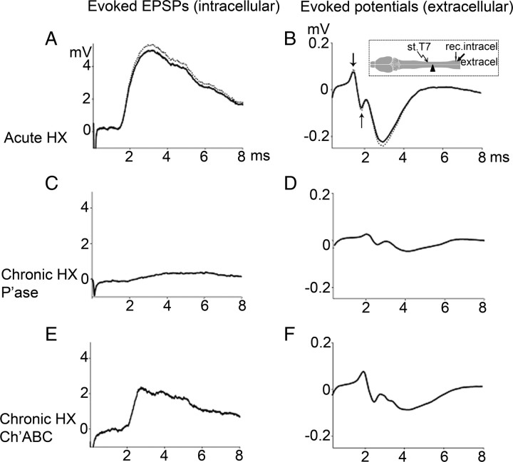
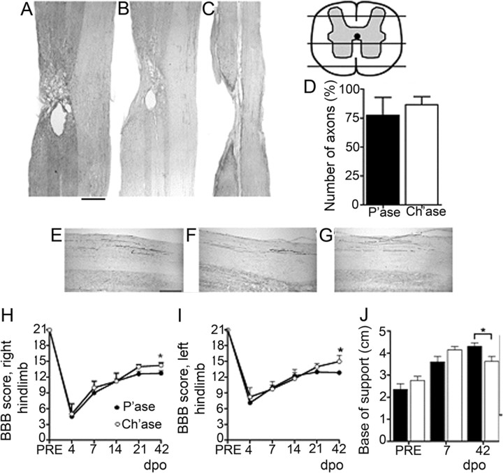
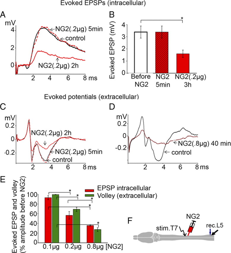
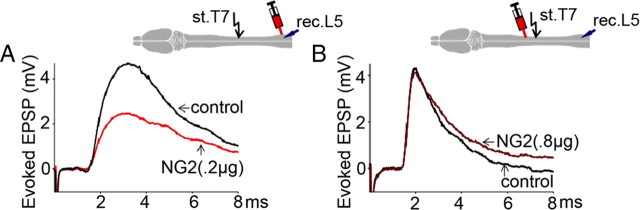
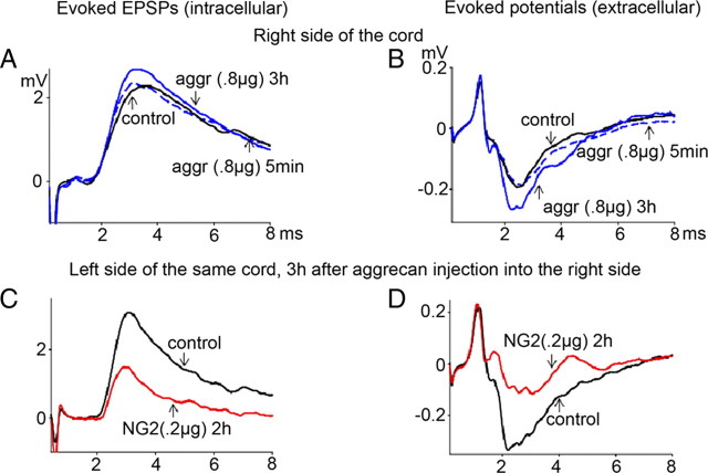
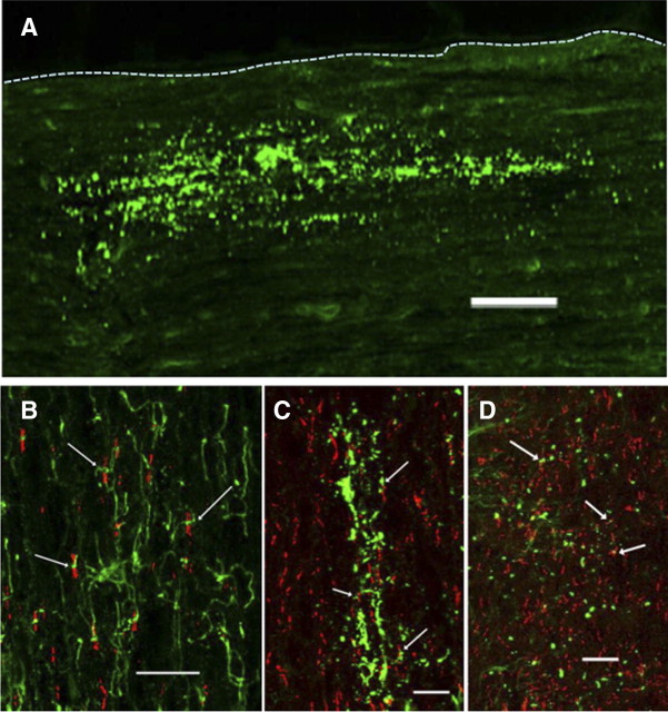
Chronic unilateral hemisection (HX) of the adult rat spinal cord diminishes conduction through intact fibers in the ventrolateral funiculus (VLF) contralateral to HX. This is associated with a partial loss of myelination from fibers in the VLF (Arvanian et al., 2009). Here, we again measured conduction through the VLF using electrical stimulation while recording the resulting volley and synaptic potentials in target motoneurons. We found that intraspinal injection of chondroitinase-ABC, known to digest chondroitin sulfate proteoglycans (CSPGs), prevented the decline of axonal conduction through intact VLF fibers across from chronic T10 HX. Chondroitinase treatment was also associated with behavior suggestive of an improvement of locomotor function after chronic HX. To further study the role of CSPGs in axonal conduction, we injected three purified CSPGs, NG2 and neurocan, which increase in the vicinity of a spinal injury, and aggrecan, which decreases, into the lateral column of the uninjured cord at T10 in separate experiments. Intraspinal injection of NG2 acutely depressed axonal conduction through the injected region in a dose-dependent manner. Similar injections of saline, aggrecan, or neurocan had no significant effect. Immunofluorescence staining experiments revealed the presence of endogenous and exogenous NG2 at some nodes of Ranvier. These results identify a novel acute action of CSPGs on axonal conduction in the spinal cord and suggest that antagonism of proteoglycans reverses or prevents the decline of axonal conduction, in addition to stimulating axonal growth.


Figures

Figure 1.
Figure 1.
Representative traces of EPSPs [intracellular (intracel)] (A, C, E) and evoked potentials [extracellular (extracel)] (B, D, F) under the following conditions: before (dotted line traces) and after (solid line traces) acute HX (A, B); chronic HX and P'ase treatment (C, D); chronic HX and Ch'ase treatment (E, F). Inset (B) shows position of stimulation (st)/recording (rec.) electrodes with respect to HX. The extracellular responses consist of two components: the early biphasic arriving volley (measured between the arrows) and the later downward extracellular synaptic response (measured from baseline to peak). Note that P'ase did not reverse the effects of chronic hemisection in attenuating the contralateral volley, whereas Ch'ase did partially attenuate it (see Results).
Figure 2.
Figure 2.
Effects of Ch'ase on the number of surviving reticulospinal axons and behavioral performance after chronic HX. A–C, Longitudinal sections at the site of injury showing the complete transection of the dorsolateral (A), lateral (B), and ventral (C) funiculi of the left spinal cord and the preservation of the right white matter tracts. BDA was injected in the right reticular formation of the animals, and labeled axons were counted in the right white matter contralateral to the injury, 1 cm above and below. Scale bar: (in A) A–C, 1000 μm. D, Summary of results demonstrating no difference in the preservation of these axons between the groups (p = 0.4). E–G, Representative images of reticulospinal axons in the contralateral white matter tracts rostral to HX (E), across from HX (F), and below the HX (G). Scale bar: (in E) E–G, 200 μm. H–J, The animal's ground locomotion performance 42 d after operation (dpo): right hindlimb BBB score (*p = 0.02) (H); left hindlimb BBB score (*p = 0.03) (I); and base of support derived from gait analysis (*p = 0.02) (J). PRE, Preoperation. Animals treated with Ch'ase showed better interlimb coordination.
Figure 3.
Figure 3.
Effects of intraspinal NG2 injections in lateral T10 on the evoked EPSP (intracellular) and the evoked composite potentials (extracellular). A, Superimposed averaged responses: control EPSP (black); 5 min after NG2 injection (dotted red); 3 h after NG2 injections (solid red). B, Summary of results demonstrating delayed depression of evoked EPSPs after NG2. All means represent peak amplitude of the responses from 7 rats, and for each rat the response was an average of maximum responses recorded intracellularly from 7–10 motoneurons before, 5 min after, and 3 h after injections of NG2 (0.2 μg). C, Evoked composite potentials in the same rat under the same conditions as in A to show depression of both action potential volley and synaptic components of evoked response after 0.2 μg of NG2. D, Higher dose of NG2 (0.8 μg) induces greater depression of the action potential volley and synaptic components of evoked composite responses; averaged responses recorded before (black) and 40 min after 0.8 μg NG2 injection (dark red). E, Summary of results demonstrating dose-dependent action of NG2 on EPSPs (intracellular, red columns) and action potential volley (extracellular, green columns). Ordinate represents mean ratio of the peak amplitudes of the maximum responses from 5–7 rats (7–10 motoneurons in each rat) before and 3 h after NG2 injections at denoted doses. F, Diagram to show the position of the stimulation (stim.)/recording(rec.)/injection electrodes. Asterisk (*) above the brackets (B, E) represents significant difference between the corresponding bars (B, p = 0.01; E, p = <0.001).
Figure 4.
Figure 4.
A, Injection of NG2 (0.2 μg) close to the recording (rec.) electrode induced depression of the evoked EPSPs (p = 0.008, n = 4) to a similar extent as an injection of NG2 in T10. B, Injection of NG2 at even higher doses (0.8 μg) rostral to the stimulation (st.) electrode had no effect (p = 0.6, n = 3).
Figure 5.
Figure 5.
Intraspinal injections of aggrecan (aggr) or neurocan (both 0.8 μg; right T10) had no effect on evoked EPSPs (intracellular) and no effect on the evoked composite potentials (extracellular). A, B, Control response (black; position of recording/injection electrodes on the right side, same as described for NG2 in Fig. 3F), 5 min after aggrecan injection (dotted blue), and 3 h after aggrecan injections (solid blue). C, D, In rats initially injected with aggrecan, subsequent contralateral injections of NG2 in contralateral white matter (stimulate left T7, recorded from left L5, 0.2 μg of NG2 injected in left T10) depressed both the EPSP (recorded intracellularly) and composite responses (recorded extracellularly) (n = 3; see Results for statistical significance).
Figure 6.
Figure 6.
Localization of endogenous and exogenous NG2 in the rat spinal cord. Adult rats were injected with NG2 (1 μg) and killed 15 or 60 min later. A, Overview of the distribution of exogenous NG2 15 min after injection. The injected protein distributes over a wide area in a linear pattern, suggesting an association with axons. Dotted line outlines the lateral edge of the cord. Scale bar, 100 μm. B, Appearance of NG2-positive oligodendrocyte progenitor cells (green) and CASPR (red) in the spinal cord contralateral to the injection site. The arrows point to areas were NG2-postive processes make close contact with nodes of Ranvier. C, D, Exogenous NG2 15 min (C) and 60 min (D) after injection. The protein is distributed in a linear fashion and the arrows point to NG2 accumulations close to CASPR-positive nodes of Ranvier. Scale bars: B–D, 20 μm.

Similar articles

Cited by

References

    1. Arvanian VL, Mendell LM. Removal of NMDA receptor Mg2+ block extends the action of NT-3 on synaptic transmission in neonatal rat motoneurons. J Neurophys. 2001;86:123–129. - PubMed
    1. Arvanian VL, Schnell L, Lou L, Golshani R, Hunanyan A, Ghosh A, Pearse DD, Robinson JK, Schwab ME, Fawcett JW, Mendell LM. Chronic spinal hemisection in rats induces a progressive decline in transmission in uninjured fibers to motoneurons. Exp Neurol. 2009;216:471–480. - PMC - PubMed
    1. Basso MD, Beattie MS, Bresnahan JC. A sensitive and reliable locomotor rating scale for open field testing in rats. J Neurotrauma. 1995;12:1–21. - PubMed
    1. Bekku Y, Rauch U, Ninomiya Y, Oohashi T. Brevican distinctively assembles extracellular components at the large diameter nodes of Ranvier in the CNS. J Neurochem. 2009;108:1266–1276. - PubMed
    1. Black JA, Waxman SG, Smith KJ. Remyelination of dorsal column axons by endogenous Schwann cells restores the normal pattern of Nav1.6 and Kv1.2 at nodes of Ranvier. Brain. 2006;129:1319–1329. - PubMed

Publication types

MeSH terms