. 2010 Aug;84(16):8141-52.
doi: 10.1128/JVI.00749-10. Epub 2010 Jun 9.

Selective induction of host genes by MVA-B, a candidate vaccine against HIV/AIDS

Affiliations

Selective induction of host genes by MVA-B, a candidate vaccine against HIV/AIDS

Susana Guerra et al. J Virol. 2010 Aug.

Abstract

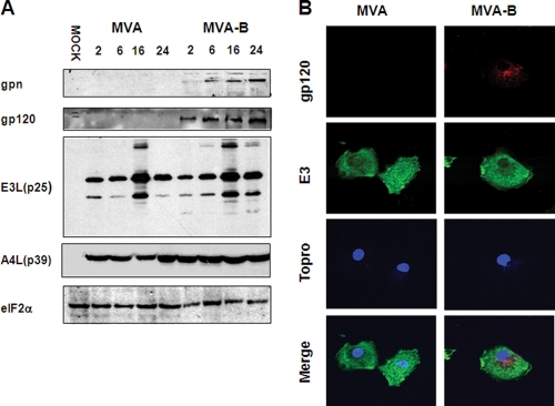
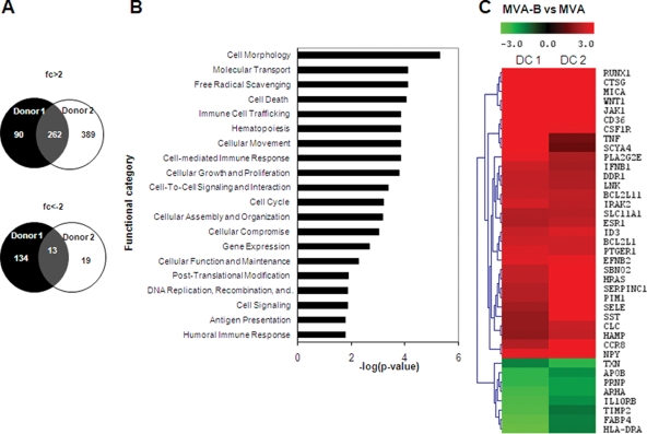
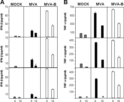
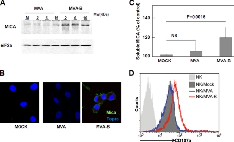
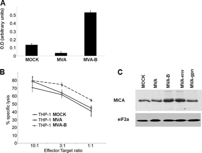
The aim of this study was to define the effects on antigen-presenting cells of the expression of HIV antigens from an attenuated poxvirus vector. We have analyzed the transcriptional changes in gene expression following infection of human immature monocyte-derived dendritic cells (DC) with recombinant modified vaccinia virus Ankara (MVA) expressing the genes encoding the gp120 and Gag-Pol-Nef antigens of HIV type 1 clade B (referred to as MVA-B) versus parental MVA infection. Using microarray technology and real-time reverse transcription-PCR, we demonstrated that the HIV proteins induced the expression of cytokines, cytokine receptors, chemokines, chemokine receptors, and molecules involved in antigen uptake and processing, including major histocompatibility complex (MHC) genes. Levels of mRNAs for interleukin-1, beta interferon, CCR8, and SCYA20 were higher after HIV antigen production. MVA-B infection also modulated the expression of antigen processing and presentation genes: the gene for MICA was upregulated, whereas those for HLA-DRA and HSPA5 were downregulated. Indeed, the increased expression of the gene for MICA, a glycoprotein related to major histocompatibility complex class I molecules, was shown to enhance the interaction between MVA-B-infected target cells and cytotoxic lymphocytes. The expression profiles of the genes for protein kinases such as JAK1 and IRAK2 were activated after HIV antigen expression. Several genes included in the JAK-STAT and mitogen-activated protein kinase signaling pathways were regulated after HIV antigen expression. Our findings provide the first gene signatures in DC of a candidate MVA-B vaccine expressing four HIV antigens and identified the biological roles of some of the regulatory genes, like that for MICA, which will help in the design of more effective MVA-derived vaccines.


Figures

FIG. 1.
FIG. 1.
Infectivity and protein synthesis evaluation in IMDDC after MVA or MVA-B infection. (A) HIV antigens and VACV protein expression in MVA- and MVA-B-infected IMDDC were detected by Western blot assay. At the times (hours) postinfection indicated above the lanes, equal amounts of proteins from extracts of cells mock infected (M) or infected with MVA or MVA-B (10 PFU/cell) were fractionated by SDS-PAGE, transferred to nitrocellulose, and incubated with antibodies against the HIV gag, HIV envelope (gp120), and specific VACV E3 early (p25) and A4 early-late (p39) proteins. eIF2α was used as a protein loading control. gpn represents the HIV-1 150-kDa fusion polyprotein Gag-Pol-Nef (B). IMDDC cultured on coverslips treated as in panel A were fixed, permeabilized, and incubated with polyclonal gp120 antibody to show the HIV envelope protein, with antibody against the VACV E3 protein, and with To-Pro to show the DNA.
FIG. 2.
FIG. 2.
Microarray analysis of HIV antigen-regulated genes in IMDDC. (A) Venn diagrams show the numbers of common or specific genes upregulated (>2-fold change) or downregulated (≤2-fold change) in MVA-B-infected IMDDC compared to MVA-infected IMDDC from two different healthy donors. A change of 2-fold indicates that gene expression in MVA-B-infected IMDDC is twice that in MVA-infected IMDDC. (B) The y axis shows the most representative high-level functions associated with genes regulated in MVA-B-infected IMDDC versus MVA-infected IMDDC, according to Ingenuity Pathway Analysis. The x axis represents the statistical significance of a given function (Fisher's exact test P value). (C) Effect of HIV-1 clade B antigen expression from MVA on host cell gene transcription. A hierarchical clustering classification of the immune response-related genes regulated in MVA-B-infected versus MVA-infected IMDDC from two donors is shown.
FIG. 3.
FIG. 3.
Validation of microarray data at the protein level. (A and B) IFN-β and TNF-α levels in cell supernatants after MVA or MVA-B infection. IMDDC from three different donors were mock infected or infected with MVA or MVA-B (10 PFU/cell), and IFN-β and TNF-α levels in supernatants were measured by ELISA. Values indicate duplicate samples in two independent experiments. An asterisk indicates a lack of data.
FIG. 4.
FIG. 4.
MICA protein upregulation after MVA-B infection correlates with activation of cytotoxic NK cells. (A) MICA levels in virus-infected IMDDC. Mock-infected (M), MVA-infected, or MVA-B-infected (10 PFU/cell) IMDDC extracts were fractionated by SDS-PAGE, transferred to nitrocellulose, and incubated with antibodies to MICA or eIF2α (used as a protein loading control). MW, molecular mass. Postinfection times (hours) are shown above the lanes. (B) Immunofluorescence analysis. IMDDC were infected with MVA or MVA-B (10 PFU/cell), and at 16 hpi, cells were fixed, permeabilized, and incubated with polyclonal MICA antibody and To-Pro to show the DNA. (C) MICA levels in cell supernatants after MVA or MVA-B infection. IMDDC from seven donors were mock infected or infected with MVA or MVA-B (10 PFU/cell), and MICA levels in supernatants were measured by ELISA. Data are expressed as percentages of the mock-infected control value. MICA shedding from MVA-B-infected IMDDC was significantly (P < 0.0015) different from that in control and MVA-infected cells, whereas the level of MICA shed by MVA-infected cells was not significantly (NS) different from that of the control. (D) Enhanced degranulation of NK cells cultured with MVA-B-infected IMDDC. Primary human NK cells maintained in IL-2 were cocultured with mock-infected DC or DC infected with either MVA or MVA-B at a ratio of two IMDDC per NK cell for 5 h. During the stimulation period, CD107a MAbs were added. After the 5-h incubation period, NK cells were identified by staining for CD56 and the level of CD107a mobilization was measured by flow cytometry. The data are representative of two independent experiments. Open dashed line, isotype control; bold continuous line, CD107a expression on NK cells cultured with mock-infected DC. Light-gray-shaded histogram, CD107a expression on NK cells cultured with MVA-infected DC; dark-gray-shaded histogram, CD107a expression on NK cells cultured with MVA-B-infected DC.
FIG. 5.
FIG. 5.
MICA protein upregulation after MVA-B infection of human THP-1 monocytes and role of gp120. (A) MVA-B infection is associated with increased levels of soluble MICA compared to those in uninfected or MVA-infected THP-1 cells. The data are representative of three independent experiments. O.D, optical density. (B) MVA-B infection leads to an increased susceptibility to NK cell lysis compared to that of uninfected and MVA-infected THP-1 cells. Primary human NK cells maintained in IL-2 were cocultured at the indicated E/T ratios with mock-infected THP-1 cells or THP-1 cells infected with either MVA or MVA-B. (C) HIV envelope increases MICA protein levels. At 6 hpi, equal amounts of proteins from cell extracts of THP-1 cells mock infected (M) or infected with MVA or MVA-B (10 PFU/cell) were fractionated by SDS-PAGE, transferred to nitrocellulose, and incubated with antibodies to MICA or eIF2α (used as a protein loading control).

Similar articles

Cited by

References

    1. Albert, M. L., M. Jegathesan, and R. B. Darnell. 2001. Dendritic cell maturation is required for the cross-tolerization of CD8+ T cells. Nat. Immunol. 2:1010-1017. - PubMed
    1. Alcamí, A., J. A. Symons, P. D. Collins, T. J. Williams, and G. L. Smith. 1998. Blockade of chemokine activity by a soluble chemokine binding protein from vaccinia virus. J. Immunol. 160:624-633. - PubMed
    1. Reference deleted.
    1. Antoine, G., F. Scheiflinger, F. Dorner, and F. G. Falkner. 1998. The complete genomic sequence of the modified vaccinia Ankara strain: comparison with other orthopoxviruses. Virology 244:365-396. - PubMed
    1. Banchereau, J., and R. M. Steinman. 1998. Dendritic cells and the control of immunity. Nature 392:245-252. - PubMed

Publication types

MeSH terms

Substances