. 2010 Aug;84(16):8085-97.
doi: 10.1128/JVI.02125-09. Epub 2010 Jun 9.

Divergent evolution of norovirus GII/4 by genome recombination from May 2006 to February 2009 in Japan

Collaborators, Affiliations

Divergent evolution of norovirus GII/4 by genome recombination from May 2006 to February 2009 in Japan

Kazushi Motomura et al. J Virol. 2010 Aug.

Abstract

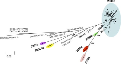
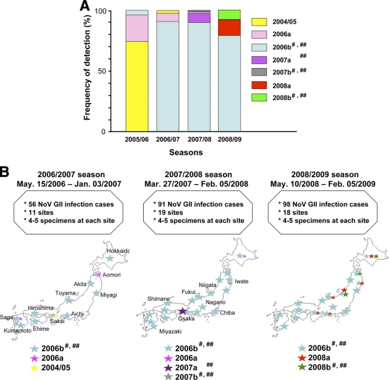
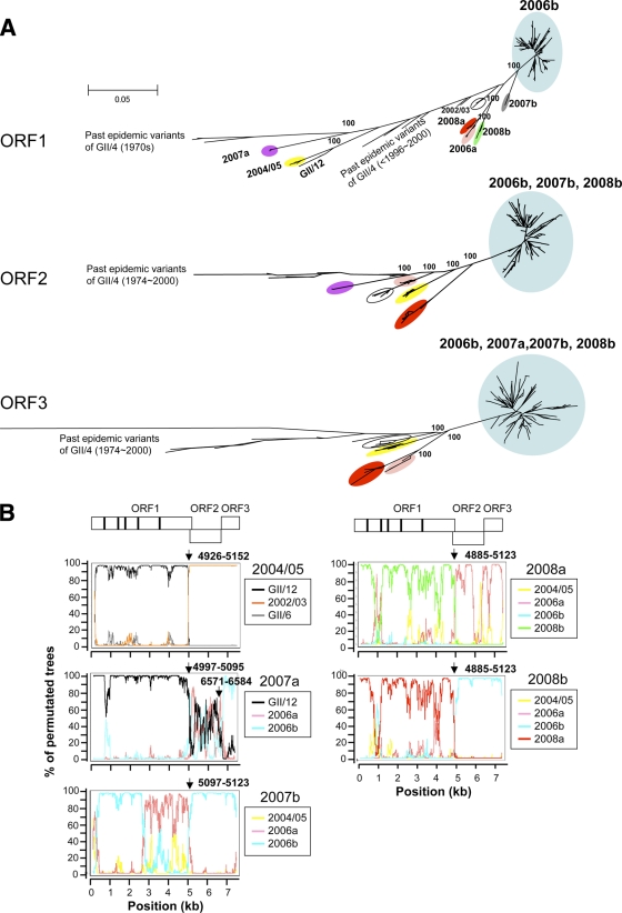
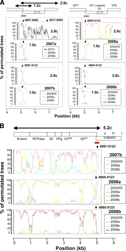
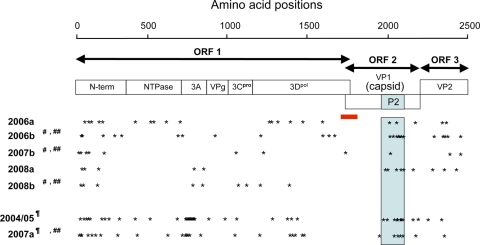
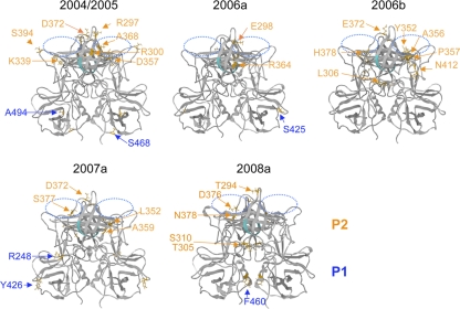
Norovirus GII/4 is a leading cause of acute viral gastroenteritis in humans. We examined here how the GII/4 virus evolves to generate and sustain new epidemics in humans, using 199 near-full-length GII/4 genome sequences and 11 genome segment clones from human stool specimens collected at 19 sites in Japan between May 2006 and February 2009. Phylogenetic studies demonstrated outbreaks of 7 monophyletic GII/4 subtypes, among which a single subtype, termed 2006b, had continually predominated. Phylogenetic-tree, bootscanning-plot, and informative-site analyses revealed that 4 of the 7 GII/4 subtypes were mosaics of recently prevalent GII/4 subtypes and 1 was made up of the GII/4 and GII/12 genotypes. Notably, single putative recombination breakpoints with the highest statistical significance were constantly located around the border of open reading frame 1 (ORF1) and ORF2 (P <or= 0.000001), suggesting outgrowth of specific recombinant viruses in the outbreaks. The GII/4 subtypes had many unique amino acids at the time of their outbreaks, especially in the N-term, 3A-like, and capsid proteins. Unique amino acids in the capsids were preferentially positioned on the outer surface loops of the protruding P2 domain and more abundant in the dominant subtypes. These findings suggest that intersubtype genome recombination at the ORF1/2 boundary region is a common mechanism that realizes independent and concurrent changes on the virion surface and in viral replication proteins for the persistence of norovirus GII/4 in human populations.


Figures

FIG. 1.
FIG. 1.
Phylogenetic classification of the NoV GII/4 subtypes in Japan during 2006 and 2009. The maximum-likelihood tree was constructed with the near-full-length genome sequences (about 7.5 kb) obtained from stool specimens collected at 19 sites in Japan between May 2006 and February 2009 in this study (n = 199) and GII/4 reference genome sequences from past epidemics in Japan and other countries in the <2000, 2002/2003, and 2004/2005 winter seasons (7, 38) (n = 18). The sequence clusters enclosed by colored ovals indicate the 7 monophyletic GII/4 subtypes identified in Japan in previous (38) and present studies.
FIG. 2.
FIG. 2.
Temporal and geographical distribution of the NoV GII/4 subtypes in Japan. The 199 near-full-length genome sequences were divided into 3 subgroups according to the collection periods: the 2006/2007 (May 2006 to January 2007) (n = 39), 2007/2008 (March 2007 to February 2008) (n = 78), and 2008/2009 (May 2008 to February 2009) (n = 82) seasons. For the analysis of the 2005/2006 season, published subtyping data (38, 43) were used (n = 38). (A) Frequencies of detection of particular NoV GII/4 subtypes in each season in Japan. (B) Geographic locations of the GII/4 subtype outbreaks. Colored stars indicate the locations of sample collection sites. Larger stars indicate the collection sites with greater frequencies of detection. #, ORF2s were classified as the same phylogenetic group (see Fig. 3A, ORF2). ##, ORF3s were classified as the same phylogenetic group (see Fig. 3A, ORF3).
FIG. 3.
FIG. 3.
Phylogenetic evidence for NoV genome mosaicism. (A) Maximum-likelihood trees of the nucleotide sequences of the complete ORF1 (about 5.1 kb), ORF2 (about 1.6 kb), and ORF3 (about 0.8 kb). The trees were constructed with the sequences obtained in previous (38) and present studies (n = 199) and the reference sequences described in Fig. 1. The GII/12 sequence (Saitama_U1/JP [25]) was used as an outgroup sequence in each tree but is shown only in the ORF1 tree. In the ORF2 and ORF3 trees, the GII/12 sequence was located far apart from the GII/4 cluster and is not shown for simplicity. (B) Bootscanning plots of nucleotide sequences of near-full-length NoV genomes. A query genome sequence (2004/05, 2007a, 2007b, 2008a, or 2008b) was aligned with three reference sequences, two sequences that were positioned relatively closely to the query sequence in the neighbor-joining trees and a sequence that was distantly related to the query sequence, using CLUSTAL W software, version 1.4 (62). The bootstrap values are plotted for a window of 300 bp moving in increments of 10 bp along the alignment using the program Simplot (48). Informative-site analyses (50) were performed using the same query and reference sequence set. Arrows indicate putative recombination breakpoints with the highest statistical significance (P ≤ 0.000001) in the informative-site analysis.
FIG. 4.
FIG. 4.
Isolation of NoV mosaic genome segments. Three genome segments (5.2, 1.0, and 2.8 kb) were amplified from the 2007a, 2007b, 2008a, and 2008b stool specimens, cloned into plasmid vectors, and sequenced. Nucleotide sequences of the cloned segments were subjected to the bootscanning-plot analysis using the same sets of reference sequences described in Fig. 3B, and the putative recombination breakpoints were assessed by informative-site analysis. (A) Results for the 2.8-kb and 1.0-kb genome segment clones (2.8c and 1.0c). (B) Results for the 5.2 kb-genome segment clones (5.2c). Red bars indicate the ORF1/ORF2 bordering region. Arrows indicate the putative recombination breakpoints with the highest statistical significance (P ≤ 0.000001) in the informative-site analysis.
FIG. 5.
FIG. 5.
Amino acid signatures of the NoV GII/4 subtypes. The deduced amino acid sequences of ORF1, ORF2, and ORF3 of a given GII/4 subtype were aligned with the GII/4 sequences identified before the outbreak season of the subtype. Amino acids specific to each subtype at the time of its first outbreak season were extracted and referred to as amino acid signatures of the new epidemic subtype. Asterisks illustrate approximate locations of the amino acid signatures in ORF1, ORF2, and ORF3. A light-blue box denotes approximate locations of the capsid P2 domain in ORF2. A red bar indicates the ORF1/2 boundary region where the single putative recombination breakpoint was assigned for each subtype genome by informative-site analyses (50). ¶, ORF1s were similar to those for GII/12 (see Fig. 4, ORF2). 2004/05 and 2007a had 27 and 63 amino acid substitutions, respectively, in ORF1s compared to the two available complete ORF1 sequences of GII/12 (accession numbers AB045603 and AB039775). #, ORF2s were classified as the same phylogenetic group (see Fig. 3A, ORF2). ##, ORF3s were classified as the same phylogenetic group (see Fig. 3A, ORF3).
FIG. 6.
FIG. 6.
3-D locations of the subtype-specific amino acids in the capsid P domain dimer. Structural models of the capsid P domain dimers of recent NoV subtypes were constructed by homology modeling as described previously (38). The 2007b and 2008b capsid models were not included because their ORF2s were classified as belonging to the same phylogenetic group as ORF2 of 2006b due to putative genome recombination (Fig. 3 and 4), and their capsid proteins had no signature or only a single signature in the P2 domain with 2006b (Fig. 5). Orange arrows and letters indicate locations and types of the unique amino acids in each GII/4 subtype at the time of its first outbreak season. Putative functional sites for virus entry into the cells are highlighted. Blue-dotted ovals, the fucose ring binding sites formed by the P domain dimer (8, 13); cyan chain, an RGD motif (60) on the β2 sheet of the P domain.

Similar articles

Cited by

References

    1. Ambert-Balay, K., F. Bon, F. Le Guyader, P. Pothier, and E. Kohli. 2005. Characterization of new recombinant noroviruses. J. Clin. Microbiol. 43:5179-5186. - PMC - PubMed
    1. Ando, T., J. S. Noel, and R. L. Fankhauser. 2000. Genetic classification of “Norwalk-like viruses.” J. Infect. Dis. 181(Suppl. 2):S336-S348. - PubMed
    1. Baker, D., and A. Sali. 2001. Protein structure prediction and structural genomics. Science 294:93-96. - PubMed
    1. Belliot, G., S. V. Sosnovtsev, T. Mitra, C. Hammer, M. Garfield, and K. Y. Green. 2003. In vitro proteolytic processing of the MD145 norovirus ORF1 nonstructural polyprotein yields stable precursors and products similar to those detected in calicivirus-infected cells. J. Virol. 77:10957-10974. - PMC - PubMed
    1. Bertolotti-Ciarlet, A., S. E. Crawford, A. M. Hutson, and M. K. Estes. 2003. The 3′ end of Norwalk virus mRNA contains determinants that regulate the expression and stability of the viral capsid protein VP1: a novel function for the VP2 protein. J. Virol. 77:11603-11615. - PMC - PubMed

Publication types

MeSH terms

Associated data