. 2010 Aug;84(16):7970-82.
doi: 10.1128/JVI.00447-10. Epub 2010 Jun 9.

Neutralization of human respiratory syncytial virus infectivity by antibodies and low-molecular-weight compounds targeted against the fusion glycoprotein

Affiliations

Neutralization of human respiratory syncytial virus infectivity by antibodies and low-molecular-weight compounds targeted against the fusion glycoprotein

Margarita Magro et al. J Virol. 2010 Aug.

Abstract

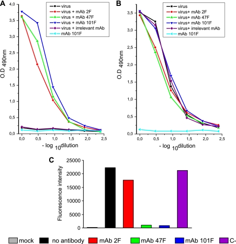
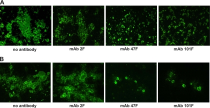
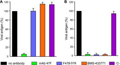
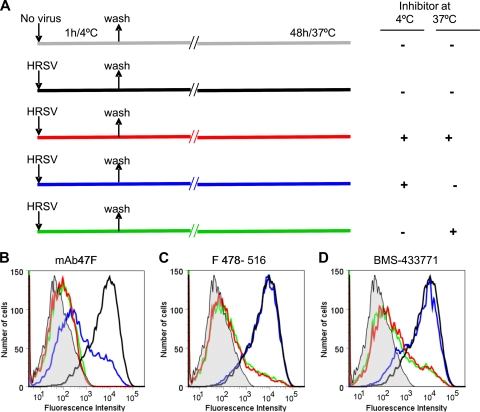
Human respiratory syncytial virus (HRSV) fusion (F) protein is an essential component of the virus envelope that mediates fusion of the viral and cell membranes, and, therefore, it is an attractive target for drug and vaccine development. Our aim was to analyze the neutralizing mechanism of anti-F antibodies in comparison with other low-molecular-weight compounds targeted against the F molecule. It was found that neutralization by anti-F antibodies is related to epitope specificity. Thus, neutralizing and nonneutralizing antibodies could bind equally well to virions and remained bound after ultracentrifugation of the virus, but only the former inhibited virus infectivity. Neutralization by antibodies correlated with inhibition of cell-cell fusion in a syncytium formation assay, but not with inhibition of virus binding to cells. In contrast, a peptide (residues 478 to 516 of F protein [F478-516]) derived from the F protein heptad repeat B (HRB) or the organic compound BMS-433771 did not interfere with virus infectivity if incubated with virus before ultracentrifugation or during adsorption of virus to cells at 4 degrees C. These inhibitors must be present during virus entry to effect HRSV neutralization. These results are best interpreted by asserting that neutralizing antibodies bind to the F protein in virions interfering with its activation for fusion. Binding of nonneutralizing antibodies is not enough to block this step. In contrast, the peptide F478-516 or BMS-433771 must bind to F protein intermediates generated during virus-cell membrane fusion, blocking further development of this process.


Figures

FIG. 1.
FIG. 1.
Antigen binding and virus neutralization with MAbs directed against the F protein of HRSV. (A) Diagram of the F protein primary structure, showing antigenic sites I, II and IV; the hydrophobic regions (▪) (the signal peptide [SP], the fusion peptide [FP], and the transmembrane region [TM]); HRA and HRB; and the sites of proteolytic processing (red arrow, site I; black arrow, site II). Shown below the diagram are the MAbs used in this study and the sequence changes in escape mutants that ablate reactivity with each MAb. (B) Serial dilutions of purified anti-F MAbs were tested in a direct ELISA for binding to a soluble form of the F protein (FTM−), as described in Materials and Methods. (C) Long virus (6.5 × 10³ PFU) was incubated with different amounts of MAbs for 30 min at 37°C before being used to infect HEp-2 cells. Production of viral antigen was quantified by ELISA 72 h later, as described in Materials and Methods, and results are presented as a percentage of the value for control cells infected in the absence of antibody. MAb 1P (against HRSV phosphoprotein) was used as a negative control in neutralization. Data represent the mean and standard deviation from three independent experiments. OD, optical density.
FIG. 2.
FIG. 2.
Neutralization with anti-F MAbs added before (A) or during (B) HRSV infection. (A) Long virus (8.2 × 10⁵ PFU) was incubated in the absence or presence of 400 μg of the anti-F MAbs indicated on the figure. Virus-antibody mixtures were ultracentrifuged (125,812 × g for 2 h in a Beckman SW60 rotor), and the pellets were resuspended and used to infect HEp-2 cells. After 72 h of incubation, production of viral antigen was quantified by ELISA, as described in Materials and Methods, and results are presented as a percentage of the value for control cells infected in the absence of antibody. MAb 1B.C11 against a capsid protein of ASPV was used as a negative control (C−) in neutralization. (B) Long virus (1 × 10⁵ PFU) ultracentrifuged as before in the absence of antibody was used to infect HEp-2 cells in the absence or presence of 3 μg of the antibodies indicated on the figure. Production of viral antigen was quantified as described for panel A. Data represent the mean and standard deviation from three independent experiments.
FIG. 3.
FIG. 3.
Quantification of antibodies, virus, and infectivity in pellets after centrifugation. Serial dilutions of pelleted virus-antibody mixtures, ultracentrifuged as described in the legend of Fig. 2A, were incubated with purified rabbit anti-FTM− antibodies bound to microtiter plates. After samples were washed, the amount of antibody bound to the captured virus was quantified with biotin-labeled anti-mouse antibodies (A), and the amount of captured virus was determined with a pool of biotin-labeled anti-F (2F, 47F, and 101F) antibodies (B), as detailed in Materials and Methods. MAb 1B.C11 against a capsid protein of ASPV was used as a negative control (C−). Controls of antibody (101F) without virus and virus without antibody were also ultracentrifuged and tested. (C) HEp-2 cells were infected with the virus-antibody mixtures present in the different pellets. After 48 h, the cells were detached, incubated with a Cy5-labeled anti-F antibody, and analyzed by flow cytometry. Mean fluorescence intensity values are shown in ordinates.
FIG. 4.
FIG. 4.
Quantification of virus binding to cells and infectivity in the presence of MAbs. (A) Long virus (6.5 × 10⁵ PFU) preincubated in the absence or presence of 30 μg of the antibodies indicated on the figure, was mixed with a suspension of HEp-2 cells for 1 h at 4°C, and the virus bound to cells was detected by flow cytometry using an anti-F Cy5 antibody. (B) Aliquots of the virus-antibody-cell mixtures were replated. Forty-eight hours postinfection, cells were resuspended and labeled with an anti-F Cy5 antibody. Mock-infected cells or cells incubated with the MAb 63G against the attachment (G) glycoprotein or with the MAb 1B.C11 (C−) against a capsid protein of ASPV were used as controls in the experiment.
FIG. 5.
FIG. 5.
Inhibition of syncytium formation by anti-F MAbs. (A) BSR-T7/5 cells were transfected with 500 ng of pTM1 plasmid encoding the full-length F gene. The transfection mixture was removed 7 h later, and the MAbs (40 μg/ml) indicated below each panel were added to the culture. Formation of syncytium was evaluated after incubation for 48 h, as described in Materials and Methods. (B) HEp-2 cells were infected with the Long strain of HRSV (multiplicity of infection, 0.1 PFU/cell). The inoculum was removed at 90 min postinfection, and cells were maintained with medium. Five hours later, MAbs were added to the culture. Syncytium formation was examined as described for panel A. The results shown in this figure are representative of three independent experiments.
FIG. 6.
FIG. 6.
Antigen binding, virus neutralization, and inhibition of syncytium formation with Fab fragments of anti-F MAbs. (A) Serial dilutions of MAbs and Fab fragments were tested for binding to a soluble form of the F protein (FTM−) in a direct ELISA, as described in Materials and Methods. (B) Long virus (6.5 × 10³ PFU) was incubated with different amounts of the indicated Fab fragments for 30 min at 37°C. Virus-Fab mixtures were then used to infect HEp-2 cells. After a 72-h incubation, production of viral antigen was quantified by ELISA, as described in Materials and Methods, and results are presented as a percentage of the value for control cells infected in the absence of antibody. Data represent the mean and standard deviation from three independent experiments. (C) HEp-2 cells were infected with the Long strain of HRSV (multiplicity of infection, 0.1 PFU/cell). The inoculum was removed at 90 min postinfection, and cells were maintained with medium. Five hours later, MAbs (40 μg/ml) or Fab fragments (1 mg/ml) were added to the culture. Formation of syncytium was evaluated after incubation for 48 h as described in Materials and Methods. The results are representative of three independent experiments.
FIG. 7.
FIG. 7.
Neutralization of HRSV by HRB F478-516 peptide and BMS-433771. (A) Schematic diagram of the F protein primary structure. The expanded region shows the amino acid sequence of the HRB F-peptide used in the study. (B) Long virus (6.5 × 10³ PFU) was incubated with increasing amounts of either HRB F478-516-derived peptide or with BMS-433771 for 30 min at 37°C before being used to infect HEp-2 cells. Production of viral antigen was quantified by ELISA 72 h later, as described in Materials and Methods, and results are presented as a percentage of the value for control cells infected in the absence of inhibitor. Data represent the mean and standard deviation from three independent experiments.
FIG. 8.
FIG. 8.
Neutralization with MAb 47F, peptide F478-516, and BMS-433771 added before (A) or during (B) HRSV infection. (A) Long virus (8.2 × 10⁵ PFU) was incubated in the absence or presence of 400 μg of MAb 47F, 925 μM F478-516, or 250 μM BMS-433771. Virus-inhibitor mixtures were ultracentrifuged (125,812 × g for 2 h in a Beckman SW60 rotor), and the pellets were resuspended and used to infect HEp-2 cells. After a 72-h incubation, production of viral antigen was quantified by ELISA as described in Materials and Methods, and results are presented as the percentage of values of infected controls without added inhibitor. MAb 1B.C11 against a capsid protein of ASPV was used as a negative control (C−) in neutralization. (B) Long virus (1 × 10⁵ PFU), ultracentrifuged as before in the absence of inhibitor, was used to infect HEp-2 cells in the absence or presence of 3 μg of MAb 47F, 7.4 μM F478-516, or 2 μM BMS-433771. Production of viral antigen was quantified as described for panel A. Data represent the mean and standard deviation from three independent experiments.
FIG. 9.
FIG. 9.
Effect of MAb 47F, peptide F418-516, and BMS-433771 during adsorption of HRSV to cells at 4°C. The experimental design is shown in panel A. Cultures of HEp-2 cells (90% confluent) were either mock infected (gray line) or infected with the Long strain of HRSV (multiplicity of infection, 0.2 PFU/cell) in the absence or presence of 80 μg of MAb 47F (B), 55 μM peptide F 478-516 (C), or 3 μM BMS-433771 (D) and incubated for 1 h at 4°C. The inoculum was subsequently removed, and after cultures were washed with DMEM-2.5% FCS, they were shifted up to 37°C either in the absence or presence of the same amounts of the corresponding inhibitor for another 48 h. At this time, cells were resuspended, and the amount of virus antigen present at the cell surface was quantified by flow cytometry as detailed in the legend of Fig. 4B.
FIG. 10.
FIG. 10.
Three-dimensional model of the HRSV F protein prefusion conformation. The model represents the proposed prefusion conformation of the HRSV F trimer, built using the SWISS-MODEL server facilities (http://swissmodel.expasy.org/) and the atomic coordinates of the prefusion structure of the PIV5 F protein (Protein Data Bank code, 2B9B) as a template (78). The backbone structure of the three monomers is shown in gray. Fusion peptide sequences of one monomer are shown in pink, and those of HRA are shown in black. Residues that are changed in virus isolates or in escape mutants selected with monoclonal antibodies, whose epitopes map in different antigenic sites of the F protein, are shown as colored spheres (antigenic site I, amino acid 389; antigenic site II, amino acids 262, 268, 272, and 275; and antigenic site IV, amino acids 429, 432, 433, 436, and 447). The two proteolytic cleavage sites are indicated with arrows in one of the monomers, colored as described in the legend of Fig. 1.

Similar articles

Cited by

References

    1. Anderson, L. J., P. Bingham, and J. C. Hierholzer. 1988. Neutralization of respiratory syncytial virus by individual and mixtures of F and G protein monoclonal antibodies. J. Virol. 62:4232-4238. - PMC - PubMed
    1. Andries, K., M. Moeremans, T. Gevers, R. Willebrords, C. Sommen, J. Lacrampe, F. Janssens, and P. R. Wyde. 2003. Substituted benzimidazoles with nanomolar activity against respiratory syncytial virus. Antiviral Res. 60:209-219. - PubMed
    1. Arbiza, J., G. Taylor, J. A. Lopez, J. Furze, S. Wyld, P. Whyte, E. J. Stott, G. Wertz, W. Sullender, M. Trudel, et al. 1992. Characterization of two antigenic sites recognized by neutralizing monoclonal antibodies directed against the fusion glycoprotein of human respiratory syncytial virus. J. Gen. Virol. 73:2225-2234. - PubMed
    1. Baker, K. A., R. E. Dutch, R. A. Lamb, and T. S. Jardetzky. 1999. Structural basis for paramyxovirus-mediated membrane fusion. Mol. Cell 3:309-319. - PubMed
    1. Beeler, J. A., and K. van Wyke Coelingh. 1989. Neutralization epitopes of the F glycoprotein of respiratory syncytial virus: effect of mutation upon fusion function. J. Virol. 63:2941-2950. - PMC - PubMed

Publication types

MeSH terms

LinkOut - more resources