

Is inclusion of Sabouraud dextrose agar essential for the laboratory diagnosis of fungal keratitis?
- PMID: 20534916
- PMCID: PMC2907027
- DOI: 10.4103/0301-4738.64122
Is inclusion of Sabouraud dextrose agar essential for the laboratory diagnosis of fungal keratitis?
Abstract
Purpose: To determine whether the inclusion of Sabouraud dextrose agar (SDA) is essential in the diagnosis of fungal keratitis.
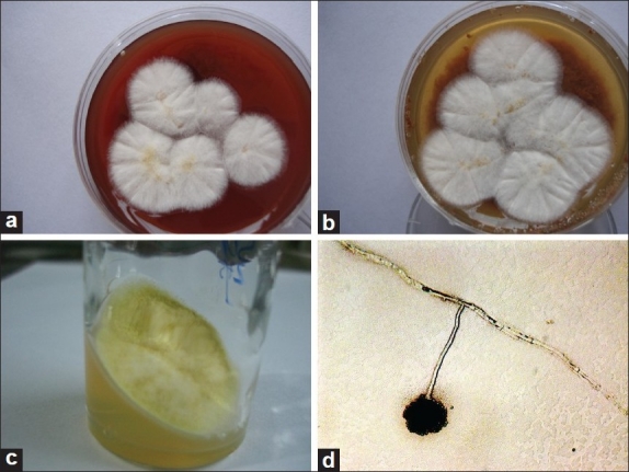
Materials and methods: Corneal scrapings of 141 patients with microbial keratitis were smeared and cultured. Sheep blood agar (BA), chocolate agar (CA), SDA, non-nutrient agar (NNA) with Escherichia coli overlay, and brain heart infusion broth (BHI) were evaluated for time taken for growth and cost. The media were also evaluated experimentally for rate of growth and time taken for identification.
Results: Twenty-six of 39 patients positive for fungus in corneal scrapings by microscopy were culture-positive. Fungus grew on BA in 22/39, on CA in 18/39, on SDA in 17/39, on NNA in 17/39, and on BHI in 13/39 cases. Growth on SDA was higher in ulcers with larger infiltrate (6/18 versus 9/13, P = 0.04). Estimated saving with inclusion of only BA/CA was Rs. 600 per patient. Performance of all media was similar in in vitro experiment although the characteristic spores and color were seen earlier on SDA.
Conclusion: Fungal keratitis can be reliably confirmed on BA or CA, which support growth of both bacteria and fungus.
Conflict of interest statement
Figures
Comment in
-
Sabouraud dextrose agar for the diagnosis of fungal keratitis.Indian J Ophthalmol. 2010 Nov-Dec;58(6):549-50; author reply 550. doi: 10.4103/0301-4738.71684. Indian J Ophthalmol. 2010. PMID: 20952848 Free PMC article. No abstract available.
-
Comment on: is inclusion of Sabouraud dextrose agar essential for the laboratory diagnosis of fungal keratitis?Indian J Ophthalmol. 2011 May-Jun;59(3):260-1. doi: 10.4103/0301-4738.81031. Indian J Ophthalmol. 2011. PMID: 21586861 Free PMC article. No abstract available.
-
Is inclusion of Sabouraud dextrose agar essential for the laboratory diagnosis of fungal keratitis?Indian J Ophthalmol. 2011 May-Jun;59(3):263-4. doi: 10.4103/0301-4738.81037. Indian J Ophthalmol. 2011. PMID: 21586864 Free PMC article. No abstract available.
References
-
- Gopinathan U, Garg P, Fernandes M, Sharma S, Athmanathan S, Rao GN. The epidemiological features and laboratory results of fungal keratitis.A 10-year review at a referral eye care centre in south India. Cornea. 2002;21:555–9. - PubMed
-
- Dunlop AA, Wright ED, Howlader SA, Nazrul I, Husain R, McClellan K, et al. Suppurative corneal ulceration in Bangladesh: A study of 142 cases, examining the microbiological diagnosis, clinical and epidemiological features of bacterial and fungal keratitis. Aust N Z J Ophthalmol. 1994;22:105–10. - PubMed
Publication types
MeSH terms
Substances
LinkOut - more resources
Full Text Sources
Medical
