

Misleading Goldmann applanation tonometry in a post-LASIK eye with interface fluid syndrome
- PMID: 20534929
- PMCID: PMC2907040
- DOI: 10.4103/0301-4738.64133
Misleading Goldmann applanation tonometry in a post-LASIK eye with interface fluid syndrome
Abstract
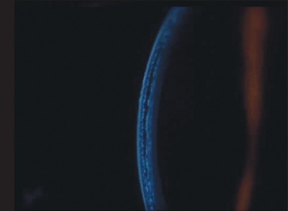
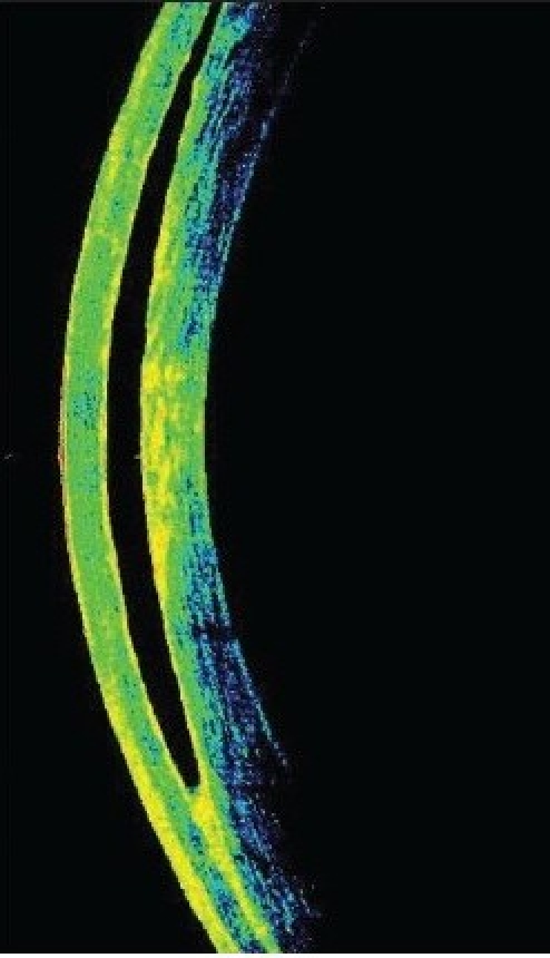
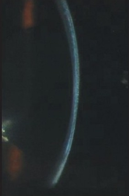
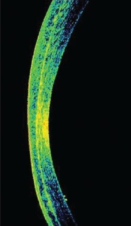
A 21-year-old myope presented with decreased vision and corneal edema following vitreoretinal surgery for retinal detachment. While intraocular pressure (IOP) measurement with Goldmann applanation tonometer (GAT) was low, the digital tonometry indicated raised pressures. An interface fluid syndrome (IFS) was suspected and confirmed by clinical exam and optical coherence tomography. A tonopen used to measure IOP through the peripheral cornea revealed elevated IOP which was the cause of the interface fluid. Treatment with IOP-lowering agents resulted in complete resolution of the interface fluid. This case is being reported to highlight the fact that IFS should be suspected when there is LASIK flap edema and IOP readings using GAT are low and that GAT is not an optimal method to measure IOP in this condition. Alternative methods like tonopen or Schiotz tonometry can be used.
Figures
Similar articles
-
Changes in biomechanical properties of the cornea and intraocular pressure after myopic laser in situ keratomileusis using a femtosecond laser for flap creation determined using ocular response analyzer and Goldmann applanation tonometry.J Glaucoma. 2015 Mar;24(3):195-201. doi: 10.1097/IJG.0b013e31829da1ec. J Glaucoma. 2015. PMID: 23807345
-
Interface Fluid Syndrome Induced by Uncontrolled Intraocular Pressure Without Triggering Factors After LASIK in a Glaucoma Patient: A Case Report.Medicine (Baltimore). 2015 Sep;94(39):e1609. doi: 10.1097/MD.0000000000001609. Medicine (Baltimore). 2015. PMID: 26426645 Free PMC article.
-
Measurement of intraocular pressure in LASIK and LASEK patients using the Reichert Ocular Response Analyzer and Goldmann applanation tonometry.J Refract Surg. 2008 Apr;24(4):366-70. doi: 10.3928/1081597X-20080401-09. J Refract Surg. 2008. PMID: 18500086
-
Finite element simulation of Goldmann tonometry after refractive surgery.Clin Biomech (Bristol). 2020 Jan;71:24-28. doi: 10.1016/j.clinbiomech.2019.09.007. Epub 2019 Oct 5. Clin Biomech (Bristol). 2020. PMID: 31677547
-
Interface fluid after laser in situ keratomileusis.J Refract Surg. 2003 Jul-Aug;19(4):455-9. doi: 10.3928/1081-597X-20030701-13. J Refract Surg. 2003. PMID: 12899478 Review.
Cited by
-
Role of AS-OCT in Managing Corneal Disorders.Diagnostics (Basel). 2022 Apr 7;12(4):918. doi: 10.3390/diagnostics12040918. Diagnostics (Basel). 2022. PMID: 35453966 Free PMC article. Review.
-
Acute interface fluid syndrome after laser in situ keratomileusis in a case of cytomegalovirus (CMV) endotheliitis and secondary glaucoma.BMJ Case Rep. 2021 Apr 30;14(4):e236742. doi: 10.1136/bcr-2020-236742. BMJ Case Rep. 2021. PMID: 33931424 Free PMC article.
-
The Effect of Corneal Refractive Surgery on Glaucoma.J Ophthalmol. 2017;2017:8914623. doi: 10.1155/2017/8914623. Epub 2017 Apr 9. J Ophthalmol. 2017. PMID: 28491472 Free PMC article. Review.
-
Limbal rebound tonometry: clinical comparisons and applications.Graefes Arch Clin Exp Ophthalmol. 2017 Sep;255(9):1795-1799. doi: 10.1007/s00417-017-3725-7. Epub 2017 Jun 28. Graefes Arch Clin Exp Ophthalmol. 2017. PMID: 28660442
-
When Goldmann applanation tonometry is not reliable in post Lasik situations.Indian J Ophthalmol. 2011 Jul-Aug;59(4):331-2. doi: 10.4103/0301-4738.82015. Indian J Ophthalmol. 2011. PMID: 21666330 Free PMC article. No abstract available.
References
-
- Lyle WA, Jin GJ. Interface fluid associated with diffuse lamellar keratitis and epithelial ingrowth after laser in situ keratomileusis. J Cataract Refract Surg. 1999;25:1009–12. - PubMed
-
- Kang SJ, Dawson DG, Hopp LM, Schmack I, Grossniklaus HE, Edelhauser HF. Interface fluid syndrome in laser in situ keratomileusis after complicated trabeculectomy. J Cataract Refract Surg. 2006;32:1560–2. - PubMed
-
- Shaikh NM, Shaikh S, Singh K, Manche E. Progression to end-stage glaucoma after laser in situ keratomileusis. J Cataract Refract Surg. 2002;28:356–9. - PubMed
-
- Hamilton DR, Manche EE, Rich LF, Maloney RK. Steroid-induced glaucoma after laser in situ keratomileusis associated with interface fluid. Ophthalmology. 2002;109:659–65. - PubMed
Publication types
MeSH terms
LinkOut - more resources
Full Text Sources
