. 2010 Jun 10;465(7299):808-12.
doi: 10.1038/nature09005.

Patient-specific induced pluripotent stem-cell-derived models of LEOPARD syndrome

Affiliations

Patient-specific induced pluripotent stem-cell-derived models of LEOPARD syndrome

Xonia Carvajal-Vergara et al. Nature. .

Abstract

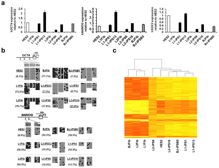
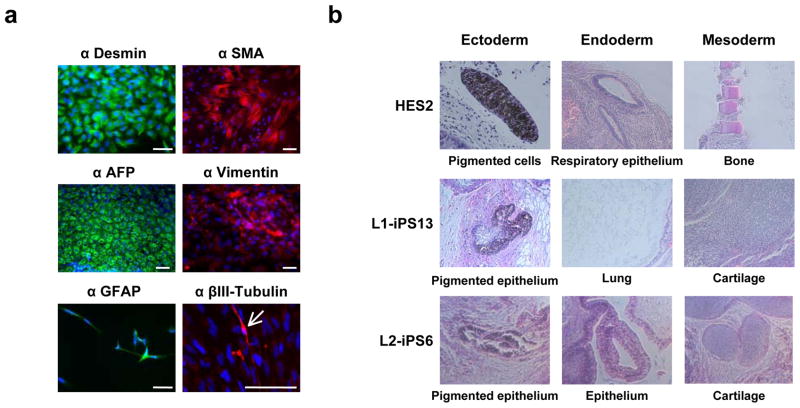
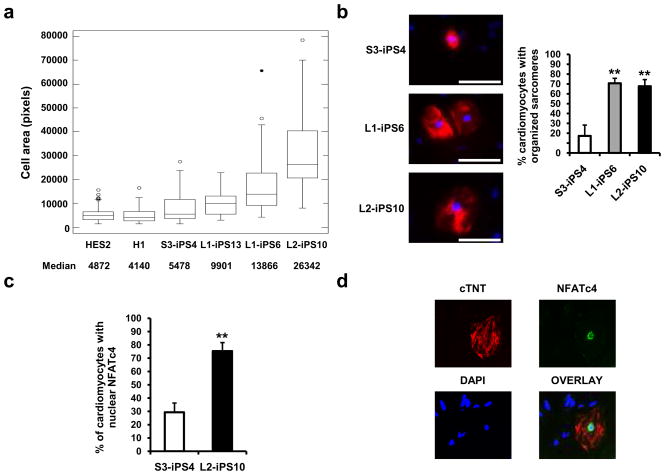
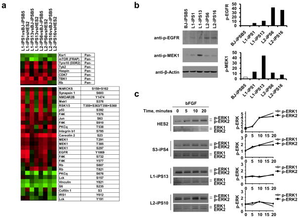
The generation of reprogrammed induced pluripotent stem cells (iPSCs) from patients with defined genetic disorders holds the promise of increased understanding of the aetiologies of complex diseases and may also facilitate the development of novel therapeutic interventions. We have generated iPSCs from patients with LEOPARD syndrome (an acronym formed from its main features; that is, lentigines, electrocardiographic abnormalities, ocular hypertelorism, pulmonary valve stenosis, abnormal genitalia, retardation of growth and deafness), an autosomal-dominant developmental disorder belonging to a relatively prevalent class of inherited RAS-mitogen-activated protein kinase signalling diseases, which also includes Noonan syndrome, with pleomorphic effects on several tissues and organ systems. The patient-derived cells have a mutation in the PTPN11 gene, which encodes the SHP2 phosphatase. The iPSCs have been extensively characterized and produce multiple differentiated cell lineages. A major disease phenotype in patients with LEOPARD syndrome is hypertrophic cardiomyopathy. We show that in vitro-derived cardiomyocytes from LEOPARD syndrome iPSCs are larger, have a higher degree of sarcomeric organization and preferential localization of NFATC4 in the nucleus when compared with cardiomyocytes derived from human embryonic stem cells or wild-type iPSCs derived from a healthy brother of one of the LEOPARD syndrome patients. These features correlate with a potential hypertrophic state. We also provide molecular insights into signalling pathways that may promote the disease phenotype.


Figures

Figure 1
Figure 1. Gene expression profile in LS-iPSC is similar to HESC
a, Quantitative real-time PCR assay for the expression of endogenous hOCT4, hNANOG and hSOX2 in iPSC and parental fibroblasts (Fib). PCR reactions were normalized against β-ACTIN and plotted relative to expression levels in HES2. Error bars indicate ± s.d. of triplicates. b, Bisulfite sequencing analyses of the OCT4 and NANOG promoters. The cell line and the percentage of methylation is indicated to the left of each cluster. c, Heat map showing hierarchical clustering of 3657 genes with at least two-fold expression change between the average of the three fibroblast cell lines versus all the iPSC lines/HES samples. Expression levels are represented by color; red indicates lower and yellow higher expression.
Figure 2
Figure 2. LS-iPSC differentiate in vitro and in vivo into all three germ layers
a, L2-iPS6 cells were differentiated as floating EBs for eight days and then plated onto gelatin-coated dishes and allowed to differentiate for another eight days. Immunocytochemistry showed cell types positively stained for differentiation markers including Desmin/SMA (mesoderm), AFP (endoderm), vimentin (mesoderm), and GFAP/βIII-Tubulin (ectoderm). The arrow indicates a βIII-tubulin-positive cell. Scale bar, 100 μm. b, HES2, L1-iPSC and L2-iPSC were injected subcutaneously into the right hindleg of immuno-compromised NOD-SCID mice. The resulting teratomas were stained with hematoxylin and eosin and tissues representative of all three germ layers were observed.
Figure 3
Figure 3. Cardiomyocytes derived from LS-iPSC show hypertrophic features
a, HES2, H1, wt S3-iPS4 and three LS-iPS clones, were differentiated into cardiac lineage. Cell areas of 50 random cTNT-positive cardiomyocytes of each cell line were measured using ImageJ. Boxes show the span from the median (50th percentile) to the first and third quartiles. The lines represent the largest/smallest sizes that are no more than 1.5 times the median to quartile distance. Additional points drawn represent extreme values. b, Sarcomeric organization was assessed in 50 cTNT positive (red) cardiomyocytes. Data are presented as mean ± s.d. n = 3; **P < 0.01 (Student’s t-test). c, S3-iPS4 and L2-iPS10 cells-derived cardiomyocytes were restained with NFATc4 antibody, and the nuclear versus cytosolic expression was analyzed. n = 3; **P < 0.01 (Student’s t-test). d, Nuclear localization of NFATc protein in a cTNT-positive cell from L2-iPS10 is shown.
Figure 4
Figure 4. Phosphoproteomic and MAPK activation analyses
a, Protein extracts of two iPSC from each LS patient (L1 and L2), wt iPSC (BJ-iPSB5) and HES2 were hybridized to an antibody microarray. The heat map represents the most significant protein changes preserved in all the comparison groups. b, pMEK1 and pEGFR expression was confirmed by Western blot using phospho-specific antibodies. Band density was measured (ImageJ software), and normalized to β-Actin. c, HES2, wt S3-iPS4, and LS-iPSC were serum- and bFGF-starved for 6 hours and then treated with bFGF (20 ng/ml) for the indicated time. Phosphorylated ERK1/2 (p-ERK1/2) and total ERK were assessed by immunoblotting and quantitated. p-ERK1/2 levels were compared to the untreated p-ERK1/2 level in each sample, normalized to the total ERK1/2 and represented graphically at the right of each panel.

Similar articles

Cited by

References

    1. Gorlin RJ, Anderson RC, Moller JH. The Leopard (multiple lentigines) syndrome revisited. Birth Defects Orig Artic Ser. 1971;07:110–115. - PubMed
    1. Sarkozy A, Digilio MC, Dallapiccola B. Leopard syndrome. Orphanet J Rare Dis. 2008;3:13. - PMC - PubMed
    1. Loh ML, et al. Mutations in PTPN11 implicate the SHP-2 phosphatase in leukemogenesis. Blood. 2004;103:2325–2331. - PubMed
    1. Tartaglia M, et al. Genetic evidence for lineage-related and differentiation stage-related contribution of somatic PTPN11 mutations to leukemogenesis in childhood acute leukemia. Blood. 2004;104:307–313. - PubMed
    1. Tartaglia M, et al. Diversity and functional consequences of germline and somatic PTPN11 mutations in human disease. Am J Hum Genet. 2006;78:279–290. - PMC - PubMed

Publication types

MeSH terms

Associated data