. 2010 Nov;17(11):1355-62.
doi: 10.1038/gt.2010.84. Epub 2010 Jun 10.

Improvement of the mdx mouse dystrophic phenotype by systemic in utero AAV8 delivery of a minidystrophin gene

Affiliations

Improvement of the mdx mouse dystrophic phenotype by systemic in utero AAV8 delivery of a minidystrophin gene

B M Koppanati et al. Gene Ther. 2010 Nov.

Abstract

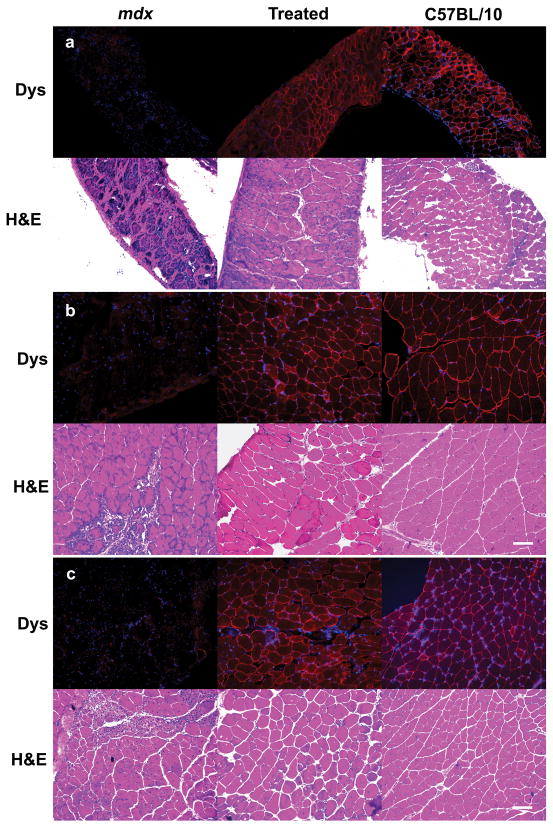
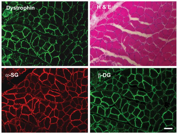
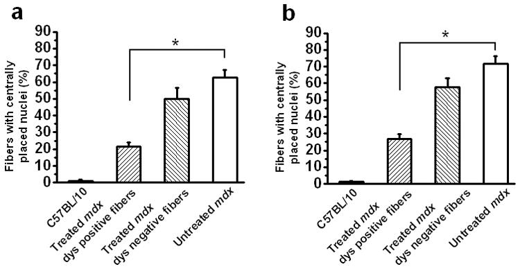
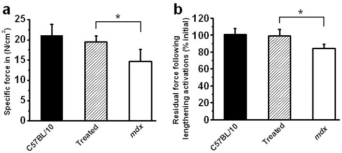
Duchenne muscular dystrophy (DMD) is a devastating primary muscle disease with pathological changes in skeletal muscle that are ongoing at the time of birth. Progressive deterioration in striated muscle function in affected individuals ultimately results in early death due to cardio-pulmonary failure. As affected individuals can be identified before birth by prenatal genetic testing for DMD, gene replacement treatment can be started in utero. This approach offers the possibility of preventing pathological changes in muscle that begin early in life. To test in utero gene transfer in the mdx mouse model of DMD, a minidystrophin gene driven by the human cytomegalovirus promoter was delivered systemically by an intraperitoneal injection to the fetus at embryonic day 16. Treated mdx mice studied at 9 weeks after birth showed widespread expression of recombinant dystrophin in skeletal muscle, restoration of the dystrophin-associated glycoprotein complex in dystrophin-expressing muscle fibers, improved muscle pathology, and functional benefit to the transduced diaphragm compared with untreated littermate controls. These results support the potential of the AAV8 vector to efficiently cross the blood vessel barrier to achieve systemic gene transfer to skeletal muscle in utero in a mouse model of muscular dystrophy, to significantly improve the dystrophic phenotype and to ameliorate the processes that lead to exhaustion of the skeletal muscle regenerative capacity.


Figures

Figure 1
Figure 1. Restoration of dystrophin and amelioration of dystrophic pathology in AAV8 minidystrophin treated mdx diaphragm, upper limb and lower limb muscles
Tissues were collected at 9 weeks after birth following an intraperitoneal injection of AAV8 minidystrophin into E-16 pups of pregnant mdx mice and analyzed for dystrophin expression. Dystrophin immunohistochemistry (upper panels) and H & E staining (lower panels) were evaluated in (a) diaphragm, (b) upper limb, and (c) lower limb muscles. Uninjected mdx littermates and C57BL/10 tissues were used as negative and positive controls, respectively, for immunohistochemistry and histology. Dys, dystrophin; α-SG, α-sarcoglycan; β-DG, β-dystroglycan. H&E, hematoxylin and eosin. Bar = 100 μm.
Figure 2
Figure 2. Restoration of dystrophin associated glycoprotein (DAG) complex proteins in lower limb muscle sections after in utero gene transfer of AAV8 minidystrophin
Gastrocnemius muscle from mice treated with AAV8 minidystrophin at E16 in utero at a dose of 6.4 × 10¹¹ vector genomes per fetus was cryosectioned. Staining of serial sections with hematoxylin and eosin (H&E) and immunohistochemistry for dystrophin, α-sarcoglycan (α-SG) and β-dystroglycan (β-DG) are shown. Bar = 100 μm.
Figure 3
Figure 3. Decreased percentage of fibers with centrally placed nuclei associated with recombinant minidystrophin expression in muscle tissues after intraperitoneal administration of AAV8 minidystrophin vector in utero
The percentage of fibers with centrally-placed nuclei was calculated from cross-sections of (a) diaphragm and (b) lower limb muscles treated by systemic in utero delivery of an AAV8 minidystrophin vector at E16 at a dose of 6.4 × 10¹¹ vector genomes per fetus. Fibers from the C57BL/10 normal mice, the untreated mdx, and the treated dystrophin positive and dystrophin negative mdx, were analyzed. The data is shown as mean ± SE. Significant differences from untreated mdx mice are shown (*P<0.05). dys, dystrophin.
Figure 4
Figure 4. Improvement in force generation properties in diaphragm after intraperitoneal administration of AAV8 minidystrophin vector in utero
The diaphragm muscles were collected at 9 weeks of age from mice treated in utero and were analyzed ex vivo. Diaphragms of control C57BL/10 (n=5 mice), AAV8 minidystrophin vector-treated mdx (n=6 mice) and untreated mdx (n=11 mice) mice were analyzed for (a) specific force (N/cm2) and (b) residual force following 10 repetitive lengthening activations divided by initial force and expressed as a percentage. The data is shown as mean ± SE. Significant differences from untreated mdx mice are shown (*P<0.05).

References

    1. Emery AE. Population frequencies of inherited neuromuscular diseases--a world survey. Neuromuscul Disord. 1991;1:19–29. - PubMed
    1. Emery AEH, Muntoni F. Duchenne Muscular Dystrophy. Oxford Univ. Press; Oxford, New York: 2003.
    1. Gregorevic P, Allen JM, Minami E, Blankinship MJ, Haraguchi M, Meuse L, et al. rAAV6-microdystrophin preserves muscle function and extends lifespan in severely dystrophic mice. Nat Med. 2006;12:787–789. - PMC - PubMed
    1. Gregorevic P, Blankinship MJ, Allen JM, Crawford RW, Meuse L, Miller DG, et al. Systemic delivery of genes to striated muscles using adeno-associated viral vectors. Nat Med. 2004;10:828–834. - PMC - PubMed
    1. Liu M, Yue Y, Harper SQ, Grange RW, Chamberlain JS, Duan D. Adeno-associated virus-mediated microdystrophin expression protects young mdx muscle from contraction-induced injury. Mol Ther. 2005;11:245–256. - PMC - PubMed

Publication types