. 2010 Jul;24(2):55-66.
doi: 10.3109/01677061003797302.

Why Drosophila to study phototransduction?

Affiliations

Why Drosophila to study phototransduction?

William L Pak. J Neurogenet. 2010 Jul.

Abstract

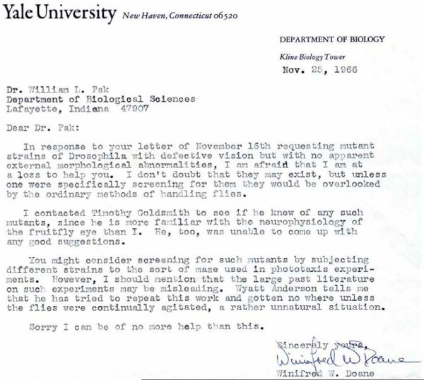
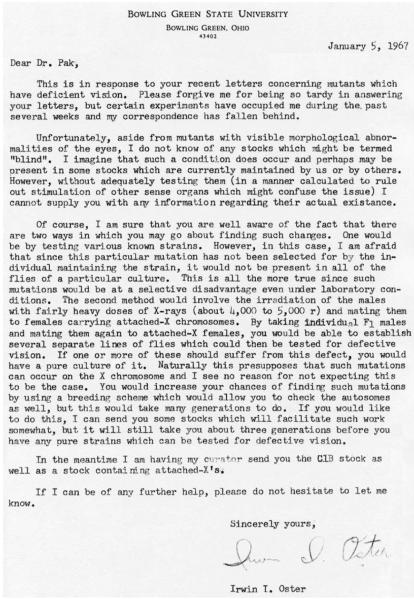
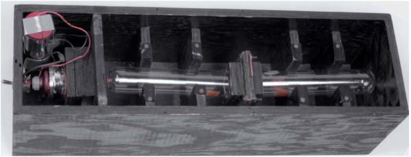
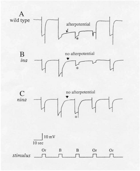
This review recounts the early history of Drosophila phototransduction genetics, covering the period between approximately 1966 to 1979. Early in this period, the author felt that there was an urgent need for a new approach in phototransduction research. Through inputs from a number of colleagues, he was led to consider isolating Drosophila mutants that are defective in the electroretinogram. Thanks to the efforts of dedicated associates and technical staff, by the end of this period, he was able to accumulate a large number of such mutants. Particularly important in this effort was the use of the mutant assay protocol based on the "prolonged depolarizing afterpotential." This collection of mutants formed the basis of the subsequent intensive investigations of the Drosophila phototransduction cascade by many investigators.


Figures

Figure 1
Figure 1
Letter from Dr. Winifred W. Doane, dated November 25, 1966. This letter was in response to the author's letter, dated November 16, 1966, inquiring if she had ever come across Drosophila mutants that acted as though blind but had no obvious external morphological defects. Reproduced with permission from Dr. Winifred W. Doane.
Figure 2
Figure 2
Letter from Dr. Irwin I. Oster, dated January 5, 1967. This letter was also in response to the same letter sent to Winifred Doane (Fig. 1) inquiring about blind mutants sent on the same day, November 16, 1966.
Figure 3
Figure 3
Phototaxis apparatus. A simple phototaxis apparatus consisted of a black box of about 17 × 17 × 56 cm in dimensions. It contained two test tubes of 1½” (3.8 cm) outside diameter and 7¾” (19.7 cm) length, placed mouth to mouth with a trap door in between. A flashlight bulb served as a light source (left compartment).
Figure 4
Figure 4
Raw phototaxis data obtained following X-ray mutagenesis. The X-rayed parent males were mated to virgin females of the attached-X stock. The F1 offspring of the above cross were tested by the “phototaxis box” (Fig. 3), and the F1 males remaining in the dark side tube (right side tube in Fig. 3) were single-male mated to virgin attached-X females. The results shown are those from the F2 offspring of this second cross, obtained on June 1 and 2, 1967. As may be seen, the phototaxis test fractionated the population of flies in each line according to their gender, almost all females going toward light and almost all males remaining in the dark. The results were consistent with the supposition that mutations induced on the X-chromosome caused the impairment of phototaxis in males. In a similar fashion, three other presumptive non-phototactic lines were isolated a week or two later.
Figure 5
Figure 5
ERG's of PDA-defective mutants, ina (inactivation but no afterpotential) (B) and nina (neither inactivation nor afterpotential) (C), compared to that of wild type (A). A bright blue stimulus elicits the prolonged depolarizing afterpotential (PDA) in wild type (arrow in A), but not in ina (arrowhead in B) or nina (arrowhead in C) mutants. A second bright blue stimulus delivered during the PDA elicits only a small response, originating from R7 and R8 photoreceptors, in wild type and ina (* in A & B). The R1-6 photoreceptors are inactivated and do not respond. By contrast, in nina mutants, R1-6 photoreceptors are not inactivated and respond with a full amplitude response to the second blue stimulus (* in C). All flies were marked with white (w) or brown;scarlet (bw;st) to eliminate the screening pigments in pigment cells. Or: orange; B: blue. Reproduced from Pak and Leung (2003) with permission from Taylor & Francis, Inc.

Similar articles

Cited by

References

    1. Ahmed ST, Joyce MV, Boggess B, O'Tousa JE. The role of Drosophila ninaG oxidoreductase in visual pigment chromophore biogenesis. J. Biol. Chem. 2006;281:9205–9209. - PubMed
    1. Alderson T. Chemically induced delayed germinal mutation in Drosophila. Nature. 1965;207:164–167. - PubMed
    1. Benzer S. Behavioral mutants of Drosophila isolated by countercurrent distribution. Proc. Natl. Acad. Sci. USA. 1967;58:1112–1119. - PMC - PubMed
    1. Bloomquist BT, Shortridge RD, Schneuwly S, Perdew M, Montell C, Steller H, et al. Isolation of a putative phospholipase C gene of Drosophila, norpA, and its role in phototransduction. Cell. 1988;54(5):723–733. - PubMed
    1. Brady AE, Limbird LE. G protein-coupled receptor interacting proteins: emerging roles in localization and signal transduction. Cell Signal. 2002;14:297–309. - PubMed

Publication types

LinkOut - more resources