

Mechanisms of excitation of spinal networks by stimulation of the ventral roots
- PMID: 20536921
- PMCID: PMC3033581
- DOI: 10.1111/j.1749-6632.2010.05535.x
Mechanisms of excitation of spinal networks by stimulation of the ventral roots
Abstract
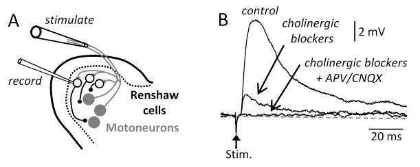
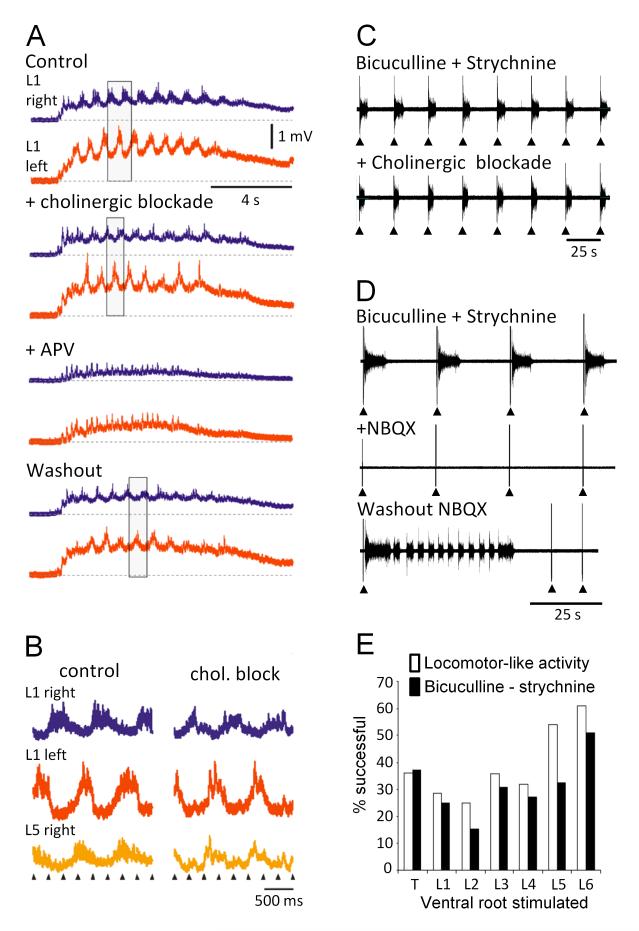
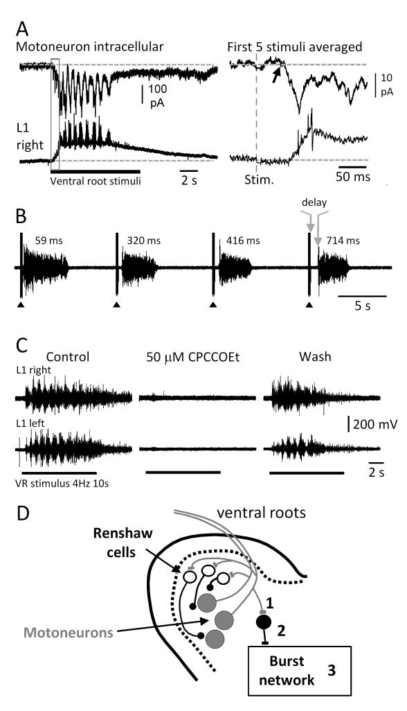
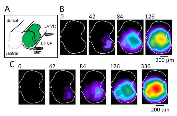
It has recently been demonstrated that motoneurons in neonatal rodents release an excitatory amino acid, in addition to acetylcholine, from their central terminals onto Renshaw cells. Although the function of this amino acid release is not understood, it may mediate the excitatory actions of motor axon stimulation on spinal motor networks. Stimulation of motor axons in the ventral roots or muscle nerves can activate the locomotor central pattern generator or entrain bursting in the disinhibited cord. Both of these effects persist in the presence of cholinergic antagonists and are abolished or diminished by ionotropic and metabotropic glutamate antagonists. Calcium imaging in the disinhibited cord shows that a ventral root stimulus evokes ventrolateral activity initially, which subsequently propagates to the rest of the cord. This finding suggests that excitatory interneurons excited by motoneuron recurrent collaterals are located in this region. However, motoneurons do not exhibit short latency excitatory potentials in response to ventral root stimulation indicating that the excitatory effects are mediated polysynaptically. We discuss the significance of these findings.
Figures
Similar articles
-
Excitatory actions of ventral root stimulation during network activity generated by the disinhibited neonatal mouse spinal cord.J Neurophysiol. 2009 Jun;101(6):2995-3011. doi: 10.1152/jn.90740.2008. Epub 2009 Mar 25. J Neurophysiol. 2009. PMID: 19321640 Free PMC article.
-
Involvement of GABA and glycine in recurrent inhibition of spinal motoneurons.J Neurophysiol. 1992 Aug;68(2):397-406. doi: 10.1152/jn.1992.68.2.397. J Neurophysiol. 1992. PMID: 1326603
-
Crossed rhythmic synaptic input to motoneurons during selective activation of the contralateral spinal locomotor network.J Neurosci. 1997 Dec 15;17(24):9433-47. doi: 10.1523/JNEUROSCI.17-24-09433.1997. J Neurosci. 1997. PMID: 9390999 Free PMC article.
-
A₁ adenosine receptor modulation of chemically and electrically evoked lumbar locomotor network activity in isolated newborn rat spinal cords.Neuroscience. 2012 Oct 11;222:191-204. doi: 10.1016/j.neuroscience.2012.07.030. Epub 2012 Jul 21. Neuroscience. 2012. PMID: 22824428
-
Positive feedback as a general mechanism for sustaining rhythmic and non-rhythmic activity.J Physiol Paris. 1995;89(4-6):241-8. doi: 10.1016/0928-4257(96)83640-0. J Physiol Paris. 1995. PMID: 8861822 Review.
Cited by
-
Balanced cholinergic modulation of spinal locomotor circuits via M2 and M3 muscarinic receptors.Sci Rep. 2019 Oct 1;9(1):14051. doi: 10.1038/s41598-019-50452-1. Sci Rep. 2019. PMID: 31575899 Free PMC article.
-
Frequency-dependent recruitment of V2a interneurons during fictive locomotion in the mouse spinal cord.Nat Commun. 2011;2:274. doi: 10.1038/ncomms1276. Nat Commun. 2011. PMID: 21505430 Free PMC article.
-
Muscarinic control of AMPA receptor responsiveness in mouse spinal cord motoneurons.J Physiol. 2012 Oct 1;590(19):4663-71. doi: 10.1113/jphysiol.2012.238444. Epub 2012 Aug 13. J Physiol. 2012. PMID: 22890702 Free PMC article.
-
Primacy of Flexor Locomotor Pattern Revealed by Ancestral Reversion of Motor Neuron Identity.Cell. 2015 Jul 16;162(2):338-350. doi: 10.1016/j.cell.2015.06.036. Cell. 2015. PMID: 26186188 Free PMC article.
-
Humans at the dawn of the in-body electrical nerve stimulation era.Facts Views Vis Obgyn. 2022 Dec;14(4):293-298. doi: 10.52054/FVVO.14.4.041. Facts Views Vis Obgyn. 2022. PMID: 36724420 Free PMC article.
References
-
- Kiehn O. Locomotor circuits in the mammalian spinal cord. Annu Rev Neurosci. 2006;29:279–306. - PubMed
-
- Perrins R, Roberts A. Cholinergic contribution to excitation in a spinal locomotor central pattern generator in Xenopus embryos. J Neurophysiol. 1995;73:1013–1019. - PubMed
-
- Wenner P, O’Donovan MJ. Mechanisms that initiate spontaneous network activity in the developing chick spinal cord. J Neurophysiol. 2001;86:1481–1498. - PubMed
Publication types
MeSH terms
Substances
Grants and funding
LinkOut - more resources
Full Text Sources
