. 2010 Jun:1198:153-8.
doi: 10.1111/j.1749-6632.2010.05501.x.

Synaptic pathways and inhibitory gates in the spinal cord dorsal horn

Affiliations

Synaptic pathways and inhibitory gates in the spinal cord dorsal horn

Tomonori Takazawa et al. Ann N Y Acad Sci. 2010 Jun.

Abstract

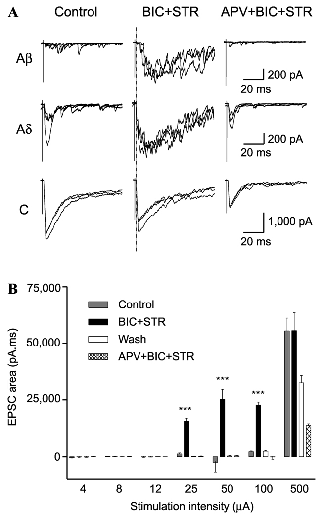
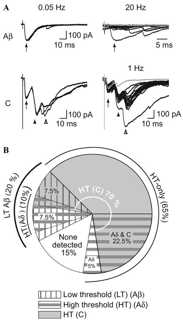
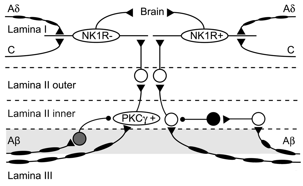
Disinhibition in the dorsal horn accompanies peripheral nerve injury and causes the development of hypersensitivity to mild stimuli. This demonstrates the critical importance of inhibition in the dorsal horn for maintaining normal sensory signaling. Here we show that disinhibition induces a novel polysynaptic low-threshold input onto lamina I output neurons, suggesting that inhibition normally suppresses a preexisting pathway that probably contributes to abnormal pain sensations such as allodynia. In addition, we show that a significant proportion of superficial dorsal horn inhibitory neurons are activated by low-threshold input. These neurons are well situated to contribute to suppressing low-threshold activation of pain output neurons in lamina I. We further discuss several aspects of inhibition in the dorsal horn that might contribute to suppressing pathological signaling.


Conflict of interest statement

Conflicts of interest

The authors declare no conflicts of interest.

Figures

Figure 1
Figure 1
Disinhibition reveals polysynaptic Aβ fiber input to lamina I NK1R+ neurons. (A, B) Show data from a neuron with C fiber monosynaptic input that has polysynaptic Aβ fiber input revealed during disinhibition. (A, left column) Example of EPSCs evoked by stimulation (0.1 ms) using Aβ (25 µA),Aδ (100 µA), and C fiber (500 µA) stimulation intensities at low frequency under control conditions. Each trace comprises three superimposed traces evoked at 0.05 Hz. (Middle column) EPSCs evoked by the same stimulation protocol but in the presence of bicuculline (BIC; 10 µM) and strychnine (STR; 300 nM). (Right column) In the presence of APV (30 µM), BIC, and STR. B, the synaptic response stimulus-intensity profile generated by calculating the total EPSC area under the curve from the artifact to the end of the recording (900 ms) for each of the three EPSCs at each intensity tested and for all conditions.
Figure 2
Figure 2
Low-threshold (Aβ) fiber input as well as input from high-threshold fibers (Aδ and/or C) to GABAergic neurons. (A) Example recordings from a P35 GABAergic neuron with input from Aβ and C fibers. (Upper panel) Three consecutive traces show responses to low-frequency Aβ fiber stimulation (left, 0.05 Hz, 25 µA, arrow). Twenty consecutive Aβ fiber responses to high-frequency stimulation (right, 20 Hz, arrow, expanded timescale). (Lower panel) C fiber input was observed when the stimulation intensities were increased (left, 0.05 Hz, 500 µA, arrowheads). The C fiber response had a monosynaptic component with no failures (but note the small amplitude in the gray trace) when tested at high frequency (right, 1 Hz, filled arrowhead). There was also a later, polysynaptic C fiber component with failures (open arrowhead, illustrated by the gray trace). (B) The proportion of GABAergic neurons with input from different afferent fiber classes is summarized. For simplicity, this representation does not distinguish between monosynaptic and polysynaptic responses. There is considerable overlap between the subsets of GABAergic neurons receiving input from each class of afferent fiber type.
Figure 3
Figure 3
Schematic diagram illustrating two putative dorsal horn neural networks involved in mechanical allodynia. The schematic diagram was drawn based on evidence reported by several groups.–,, NK1R+ neurons that receive innocuous input through polysynaptic pathways seem to be part of a local excitatory circuit that mediates mechanical allodynia after loss of GABAergic and glycinergic inhibitory control. Inhibitory interneurons in lamina II (black) receiving polysynaptic Aβ fiber input may inhibit neurons that relay non-nociceptive information to NK1R+ neurons. Moreover, PKC γ+ neurons that receive innocuous input via Aβ fibers at the lamina II/III border (shaded) are part of a local excitatory circuit that mediates dynamic mechanical allodynia after loss of glycinergic inhibitory control. We tentatively put a glycinergic inhibitory interneuron (gray) at the lamina II/III border. Given local inhibitory synaptic connection within same lamina, glycinergic inhibitory interneurons may be at lamina II/III border, although their exact location is still unknown. The target of the PKC γ+ neuron appears to be lamina I neurons lacking NK1 receptors.

References

    1. Kato G, et al. Organization of intralaminar and translaminar neuronal connectivity in the superficial spinal dorsal horn. J. Neurosci. 2009;29:5088–5099. - PMC - PubMed
    1. Miraucourt LS, Dallel R, Voisin DL. Glycine inhibitory dysfunction turns touch into pain through PKCgamma interneurons. PLoS ONE. 2007;2:e1116. - PMC - PubMed
    1. Miraucourt LS, et al. Glycine inhibitory dysfunction induces a selectively dynamic, morphine-resistant, and neurokinin 1 receptor- independent mechanical allodynia. J. Neurosci. 2009;29:2519–2527. - PMC - PubMed
    1. Torsney C, MacDermott AB. Disinhibition opens the gate to pathological pain signaling in superficial neurokinin 1 receptor-expressing neurons in rat spinal cord. J. Neurosci. 2006;26:1833–1843. - PMC - PubMed
    1. Keller AF, et al. Transformation of the output of spinal lamina I neurons after nerve injury and microglia stimulation underlying neuropathic pain. Mol. Pain. 2007;3:27. - PMC - PubMed

Publication types