Review
. 2010 Nov;21(11):1015-32.
doi: 10.1016/j.jnutbio.2010.01.005.

Liver fatty acid-binding protein and obesity

Affiliations
Review

Liver fatty acid-binding protein and obesity

Barbara P Atshaves et al. J Nutr Biochem. 2010 Nov.

Abstract

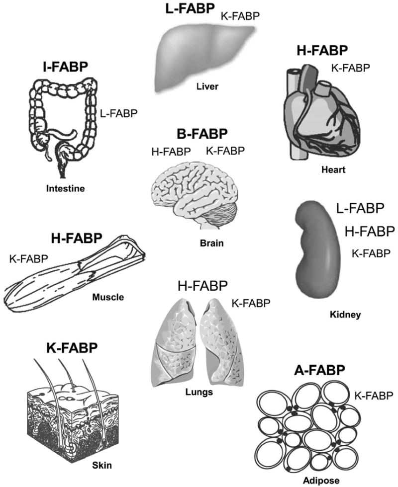
While low levels of unesterified long chain fatty acids (LCFAs) are normal metabolic intermediates of dietary and endogenous fat, LCFAs are also potent regulators of key receptors/enzymes and at high levels become toxic detergents within the cell. Elevated levels of LCFAs are associated with diabetes, obesity and metabolic syndrome. Consequently, mammals evolved fatty acid-binding proteins (FABPs) that bind/sequester these potentially toxic free fatty acids in the cytosol and present them for rapid removal in oxidative (mitochondria, peroxisomes) or storage (endoplasmic reticulum, lipid droplets) organelles. Mammals have a large (15-member) family of FABPs with multiple members occurring within a single cell type. The first described FABP, liver-FABP (L-FABP or FABP1), is expressed in very high levels (2-5% of cytosolic protein) in liver as well as in intestine and kidney. Since L-FABP facilitates uptake and metabolism of LCFAs in vitro and in cultured cells, it was expected that abnormal function or loss of L-FABP would reduce hepatic LCFA uptake/oxidation and thereby increase LCFAs available for oxidation in muscle and/or storage in adipose. This prediction was confirmed in vitro with isolated liver slices and cultured primary hepatocytes from L-FABP gene-ablated mice. Despite unaltered food consumption when fed a control diet ad libitum, the L-FABP null mice exhibited age- and sex-dependent weight gain and increased fat tissue mass. The obese phenotype was exacerbated in L-FABP null mice pair fed a high-fat diet. Taken together with other findings, these data suggest that L-FABP could have an important role in preventing age- or diet-induced obesity.


Figures

Figure 1
Figure 1. Distribution of fatty acid binding proteins (FABPs) in tissues important for long chain fatty acid (LCFA) metabolism
FABPs present in tissues at highest concentration are shown in large bold letters. FABPS present at lower concentration are shown in large unbold letters. Low expression is shown with small bold letters. The nomenclature of the long chain fatty acid binding protein family has been described [21]: L-FABP, liver type fatty acid binding protein (Fabp1 gene); I-FABP, intestinal type fatty acid binding protein (Fabp2 gene); H-FABP, heart type fatty acid binding protein (Fabp3 gene); A-FABP, adipocyte type fatty acid binding protein (Fabp4 gene); K-FABP, keratinocyte type fatty acid binding protein (also called epidermal fatty acid binding protein, E-FABP, Fabp5 gene); B-FABP, brain type fatty acid binding protein (Fabp7 gene). Additional members of the FABP family (not shown) that bind other types of ligands include: M-FABP, myelin (peripheral) type fatty acid binding protein (Fabp8 gene); T-FABP, testis type fatty acid binding protein; ILBP, ileal bile acid binding protein (Fabp6 gene); CRBP I and II, cellular retinol binding proteins I and II; and CRABP I and II, cellular retinoic acid binding proteins I and II.
Figure 2
Figure 2. Model of L-FABP functions in living cells
Bold arrows refer to reactions most greatly enhanced by L-FABP. Abbreviations are as follows: BSA, serum albumin; FA, long chain fatty acid; FATP, plasma membrane fatty acid transport protein; CD36, plasma membrane fatty acid translocase protein; CoA, coenzyme A; L-FABP, liver fatty acid binding protein; CPT-1, carnitine palmitoyl transferase I (outer mitochondrial membrane); CPT-2, carnitine palmitoyl transferase II (inner mitochondrial membrante); CAR, carnitine; G-3-P, glycerol-3-phosphate; GPAT, glycerol-3-phosphate acyltransferase; LAT, lysophosphatidic acid acyltransferase; PA, phosphatidic acid; TG, triacylglyceride; C, cholesterol; ACAT, acyl CoA cholesterol acyl transferase; CE, cholesteryl ester; VLDL very low density lipoprotein (applies only to the liver); ER, endoplasmic reticulum; PPARα, peroxisome proliferator activated receptor α; HNF4α, hepatocyte nuclear factor 4α.
Figure 3
Figure 3. Sequence homology in the liver fatty acid binding proteins (L-FABPs) from different species
(A) Amino acid sequence alignment for L-FABP derived from rat, mouse, human, and bovine. Identical amino acids are indicated by dots. (B) Promoter region of the human L-FABP gene. Response elements within the promoter for two putative sterol response elements (SRE 1 and 2), activator protein (AP1), CCAAT/enhancer binding protein (C/EBP), and the peroxisomal proliferator response element (PPRE) are indicated.
Figure 4
Figure 4. Interaction of L-FABP with carnitine palmitoyl transferase I (CPT1) determined by circular dichroism
(A) Individual far-UV circular dichroic (CD) spectra of WT CPTI C-terminal 89-residue peptide (filled circles) and an equal amino acid molarity of L-FABP (open circles). (B) Comparison of the far-UV CD spectra of an equal amino acid molarity mixture of WT CPT peptide and L-FABP obtained experimentally (actual, filled circles) and the theoretically expected spectrum (theoretical, open circles) if no conformational change occurred (i.e. the average of the two proteins). (C) Proportion of secondary structures (e.g. α–helix, β-sheet, turn, unordered) in equal molarity mixtures of CPT peptide and L-FABP obtained experimentally (actual, filled bars) and the theoretically expected (theoretical, open bars). Asterisks represent significant differences between the actual and theoretical for each compositional component; * P < 0.05; *** P < 0.001.
Figure 5
Figure 5. Correlation of LCFA oxidation with hepatic L-FABP levels in vivo
Levels of serum β-hydroxybutyrate (A) as well as L-FABP (B), SCP-2 (C), and SCP-x (D) in liver homogenates were determined in male SCP2/SCP-x null, C57BL6N wild-type, SCP-x null, and L-FABP null mice fed control standard rodent chow diet fed ad libitum.
Figure 6
Figure 6. Effect of lithogenic diet on body weight, fat tissue mass, and lean tissue mass
Average daily food consumption (A), percent change in body weight (B), fat tissue mass (C), and lean tissue mass (D) was determined in male and female mice fed a control (hatched bar) and lithogenic (solid bar) diet for 4 weeks. Values represent the mean ± SEM, n=5-7. Statistical analysis was as follows: * p≤ 0.04 vs male L-FABP +/+ on control-diet; * * p≤ 0.03 vs female L-FABP +/+ on control-diet; @ p≤ 0.004 vs male L-FABP -/- on control-diet; + p≤ 0.001 vs female L-FABP -/- on control-diet; ˆ p≤ 0.005 vs male L-FABP +/+ on lithogenic diet; # p≤ 0.004 vs male L-FABP -/- on lithogenic diet; $ p≤ 0.02 vs female L-FABP +/+ on lithogenic diet.

References

    1. Ockner RK, Manning JA, Poppenhausen RB, Ho WK. A binding protein for fatty acids in cytosol of intestinal mucosa, liver, myocardium, and other tissues. Science. 1972;177:56–58. - PubMed
    1. Glatz JF, Veerkamp JH. Intracellular fatty acid-binding proteins. [Review] Int J Biochem. 1985;17:13–22. - PubMed
    1. Glatz JFC, Van der Vusse GJ, Veerkamp JH. Fatty acid-binding proteins and their physiological significance. New Physiol Sci. 1988;3:41–43.
    1. Paulussen RJA, Veerkamp JH. Intracellular fatty acid-binding proteins characteristics and function. In: Hilderson HJ, editor. Subcellular Biochemistry. New York: Plenum Press; 1990. pp. 175–226. - PubMed
    1. Veerkamp JH, van Moerkerk HT. Fatty acid-binding protein and its relation to fatty acid oxidation. Mol Cell Biochem. 1993;123:101–106. - PubMed

Publication types