. 2010 Aug 15;344(2):784-94.
doi: 10.1016/j.ydbio.2010.05.512. Epub 2010 Jun 9.

nev (cyfip2) is required for retinal lamination and axon guidance in the zebrafish retinotectal system

Affiliations

nev (cyfip2) is required for retinal lamination and axon guidance in the zebrafish retinotectal system

Andrew J Pittman et al. Dev Biol. .

Abstract

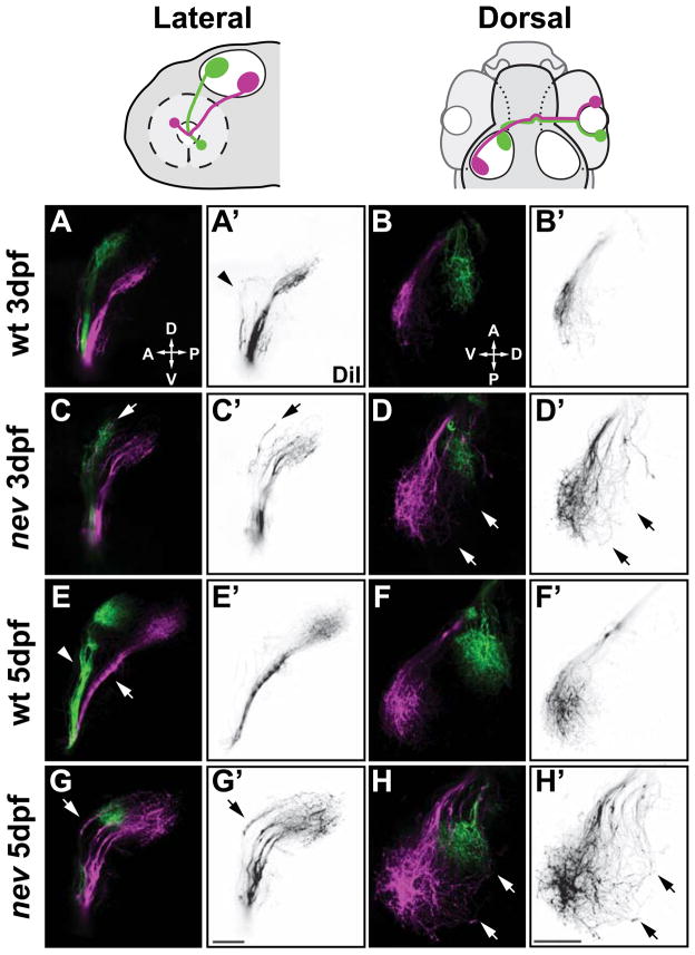
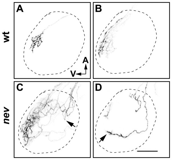
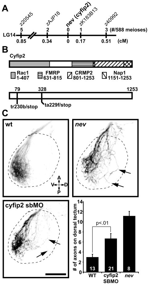
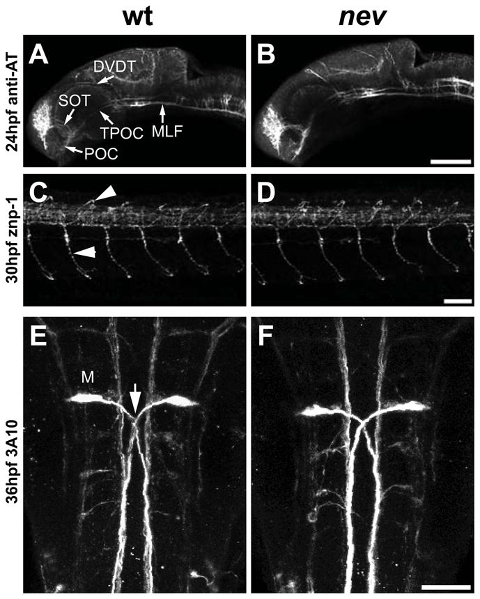
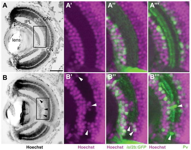
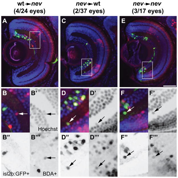
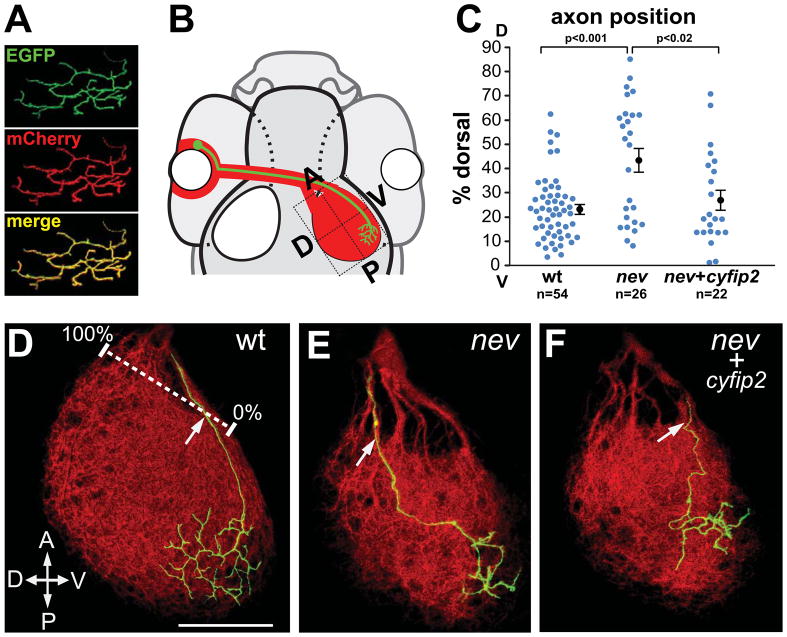
In the zebrafish retinotectal system, retinal ganglion cells (RGCs) project topographically along anterior-posterior (A-P) and dorsal-ventral (D-V) axes to innervate their primary target, the optic tectum. In the nevermind (nev) mutant, D-V positional information is not maintained by dorsonasal retinal axons as they project through the optic tract to the tectum. Here we present a detailed phenotypic analysis of the retinotectal projection in nev and show that dorsonasal axons do eventually find their correct location on the tectum, albeit after taking a circuitous path. Interestingly, nev seems to be specifically required for retinal axons but not for several non-retinal axon tracts. In addition, we find that nev is required both cell autonomously and cell nonautonomously for proper lamination of the retina. We show that nev encodes Cyfip2 (Cytoplasmic FMRP interacting protein 2) and is thus the first known mutation in a vertebrate Cyfip family member. Finally, we show that CYFIP2 acts cell autonomously in the D-V sorting of dorsonasal RGC axons in the optic tract. CYFIP2 is a highly conserved protein that lacks known domains or structural motifs but has been shown to interact with Rac and the fragile-X mental retardation protein, suggesting intriguing links to cytoskeletal dynamics and RNA regulation.


Figures

Figure 1
Figure 1
nevermind/cyfip2 is required for axon sorting and targeting of dorsonasal retinal axons. Confocal projections of dorsonasal axons (in magenta) and ventrotemporal axons (in green) in WT (A, B, E, F) and nev (C, D, G, H) at 3dpf (A–D) and 5dpf (E–H). (A′–H′) show only DiI injected dorsonasal axons in reverse contrast. Cartoons above show orientation of lateral views (A, C, E, G) and dorsal views (B, D, F, H). WT axons (A and E) from dorsonasal and ventrotemporal retina are topographically sorted in the ventral branch (arrow in E) and dorsal branch (arrowhead in E) of the optic tract, respectively. Arrowhead in A′ shows a few dorsal axons in WT projecting in the dorsal branch but turning before entering the tectum. Once on the tectum, WT axons project topographically to their target (B and F). However, in nev, dorsonasal axons are missorted in the dorsal branch of the optic tract (arrows in C and G), and project through the dorsal half of the optic tectum (arrows in D and H). Scale bars=50μm.
Figure 2
Figure 2
Dorsonasal axons take circuitous routes on the tectum in nev. (A–D) Confocal images of the left optic tectum after DiI labeling of a few RGCs in the dorsal retina of wt (A and B) or nev (C and D) at 5dpf. Dashed line indicates approximate outline of tectum. In WT, dorsonasal axons enter the tectum through the ventral branch of the optic tract, project directly to their topographic target and arborize (A and B). In nev, dorsonasal axons that enter through the dorsal branch of the optic tract meander around the dorsal tectum, sometimes turning back anteriorly after entering the tectum (arrow in C) but appear to orient toward the ventral tectum (arrow in D). Scale bar in D=50μm.
Figure 3
Figure 3
nevermind encodes Cyfip2. (A) Meiotic map of nev/cyfip2. Using a combination of SSLPs and SNPs, nev was fine-mapped to a 0.51cM interval on LG14. Numbers above show recombinations in 588 meioses; numbers below indicate distance in cM. (B) Structure of Cyfip2. Domains known to be important for protein interactions with Cyfip1 and Cyfip2 are shown (see text for references). cDNA sequencing from tr230b and ta229f mutant embryos identified premature stop codons 79 amino acids (tr230b) and 328 amino acids (ta229f) into the protein. (C) A cyfip2 splice-blocking morpholino phenocopies nev. Dorsal views showing dorsonasal axons projecting onto the optic tectum. Dashed lines indicate approximate outline of the tectum. Dorsonasal axons in nev project inappropriately onto the dorsal half of the optic tectum, as do dorsonasal axons in embryos injected with a splice-blocking morpholino (SBMO) to cyfip2 (arrows). To quantify these errors, the number of axons projecting through the dorsal optic tectum was counted in wt, cyfip2 SBMO, and nev at 3dpf. Numbers of embryos shown inside bars. Scale bar =50μm.
Figure 4
Figure 4
cyfip2 is broadly expressed in the CNS. (A) Animal pole view of a two-cell embryo showing maternal expression of cyfip2. (B–C) Lateral views of a 15 somite and 24hpf embryo, respectively. cyfip2 is broadly expressed in the CNS. (D–E) From 48hpf to 72hpf, when axons first arrive at the optic tectum and begin to innervate it, cyfip2 is still broadly expressed in the brain and retina. (F) Coronal section through a 72hpf eye. cyfip2 is expressed in the RGC layer (black arrow), the inner plexiform layer (black arrowheads), and the inner nuclear layer (white arrow). Scale bars=100μm (A–E), 50μm (F).
Figure 5
Figure 5
Early axon pathfinding appears normal in nev. Confocal projections in WT (A, C, E) and nev (B, D, F) stained with anti-acetylated tubulin (A and B), znp-1 (B and E), and 3A10 (C and F). Lateral views (A–D) and dorsal views (E and F). Anti-acetylated tubulin labels early axon pathways at 24hpf including the anterior commissure (AC), post-optic commissure (POC), supra-optic tract (SOT), tract of the post-optic commissure (TPOC), dorso-ventral diencephalic tract (DVDT), and medial longitudinal fasciculus (MLF). No differences are detectable between WT (A) and nev (B). Axon trajectories of primary motor neurons at 30hpf are normal in nev (D) compared to WT (C), including middle primary motor neurons (MiPs, arrowheads) and caudal primary motor neurons (CaPs, arrows). (E and F) Mauthner neurons and their axons (arrow in E) project normally in nev (F) as compared to WT (E) at 36hpf. Scale bar in B= 100μm, D, F= 50μm.
Figure 6
Figure 6
Retinal lamination is disrupted in nev. Coronal sections through a WT eye (A–A‴) and a nev eye (B–B‴) at 5dpf. (A and B) Hoechst 33342 stain to visualize lamination. The retinal ganglion cell (RGC) layer, inner plexiform layer (IPL), inner nuclear layer (INL), and outer nuclear layer (ONL) are all clearly visible at this stage. Arrowheads in B show displaced cells in the IPL of nev. Boxed regions are magnified in A′–A‴ and B′–B‴ and show nuclei labeled with Hoechst 33342 (magenta; A′–A‴, B′–B‴), RGCs labeled with isl2b:GFP (green; A″, B″), and a subset of amacrine cells labeled with anti-parvalbumin (green; A‴, B‴). In nev, RGCs and amacrine cells are intermingled in the IPL. Arrowheads in B″ and B‴ show displaced RGCs and amacrine cells in the IPL, respectively. ON, optic nerve. Scale bar= 50μm.
Figure 7
Figure 7
cyfip2 acts both cell autonomously and cell nonautonomously in lamination. Representative coronal sections through a nev eye with WT donor cells (A–B‴), a WT eye with nev donor cells (C–D‴), and a nev eye with nev donor cells (E–F‴). (A, C, E) Hoechst 33342 stain (blue), biotin-dextran-positive (BDA+) donor cells (red), and isl2b:GFP+ donor RGCs (green). Insets are shown magnified below. (B–B‴) WT donor cells in a nev host are misplaced in the IPL, showing nev can act cell nonautonomously. Arrows show a BDA+ donor cell next to a BDA- cell in the IPL. (D–D‴) nev donor cells in a WT host are misplaced in the IPL, showing that nev can also act cell autonomously. Arrows show a BDA+/isl2b:GFP+ cell in the IPL. (F–F‴) Control transplants show nev donor cells in a nev host that are misplaced in the IPL. Arrow shows a BDA+/isl2b:GFP+ cell misplaced in the IPL. Scale bar=50μm.
Figure 8
Figure 8
cyfip2 acts cell autonomously in dorsonasal RGC axon sorting in the optic tract. (A) Co-electroporation yields consistent co-labeling with EGFP and mCherry. (B) Diagram of rescue experiments: electroporated dorsonasal RGCs project to the posteroventral quadrant of the tectum. (C) Quantification of D–V sorting of dorsonasal axons in the optic tract as they enter the right optic tectum, relative to the the ventralmost (0%) and dorsalmost (100%) fascicles (white ruler in D). A subset of nev axons missorts into the dorsal brachium; electroporation with cyfip2 yields significant rescue. Error bars show mean ± SEM. (D–F) Confocal projections of single dorsonasal axons (GAP43-GFP, green) in the tectal neuropil (mCherryCAAX, red) at 5 dpf. (D) WT axons enter the tectum ventrally. (E) nev axons often enter dorsally. (F) cyfip2-coelectroporated nev axons are rescued and enter ventrally. Scale bar, 50 μm.

Similar articles

Cited by

References

    1. Avanesov A, Malicki J. Approaches to study neurogenesis in the zebrafish retina. Methods Cell Biol. 2004;76:333–84. - PubMed
    1. Baier H, Klostermann S, Trowe T, Karlstrom RO, Nusslein-Volhard C, Bonhoeffer F. Genetic dissection of the retinotectal projection. Development. 1996;123:415–25. - PubMed
    1. Bardoni B, Mandel JL. Advances in understanding of fragile X pathogenesis and FMRP function, and in identification of X linked mental retardation genes. Curr Opin Genet Dev. 2002;12:284–93. - PubMed
    1. Bianco IH, Carl M, Russell C, Clarke JD, Wilson SW. Brain asymmetry is encoded at the level of axon terminal morphology. Neural Dev. 2008;3:9. - PMC - PubMed
    1. Bogdan S, Grewe O, Strunk M, Mertens A, Klambt C. Sra-1 interacts with Kette and Wasp and is required for neuronal and bristle development in Drosophila. Development. 2004;131:3981–9. - PubMed

Publication types