Comparative Study
. 2010 Sep 1;169(3):1376-91.
doi: 10.1016/j.neuroscience.2010.05.081. Epub 2010 Jun 9.

The pattern of expression of guanine nucleotide-binding protein beta3 in the retina is conserved across vertebrate species

Affiliations
Comparative Study

The pattern of expression of guanine nucleotide-binding protein beta3 in the retina is conserved across vertebrate species

E R Ritchey et al. Neuroscience. .

Abstract

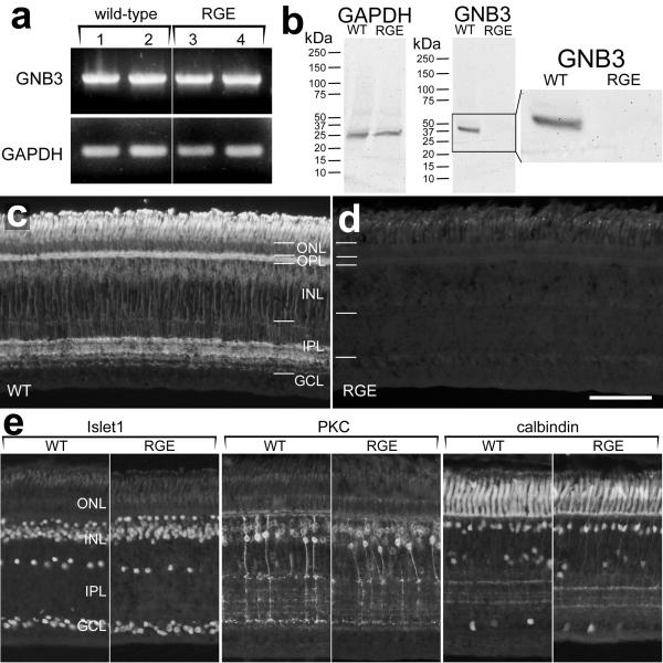
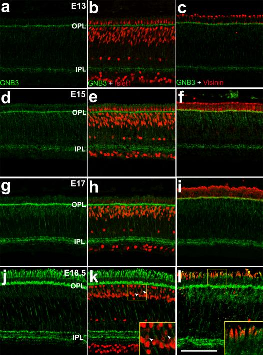
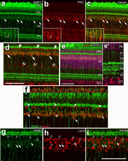
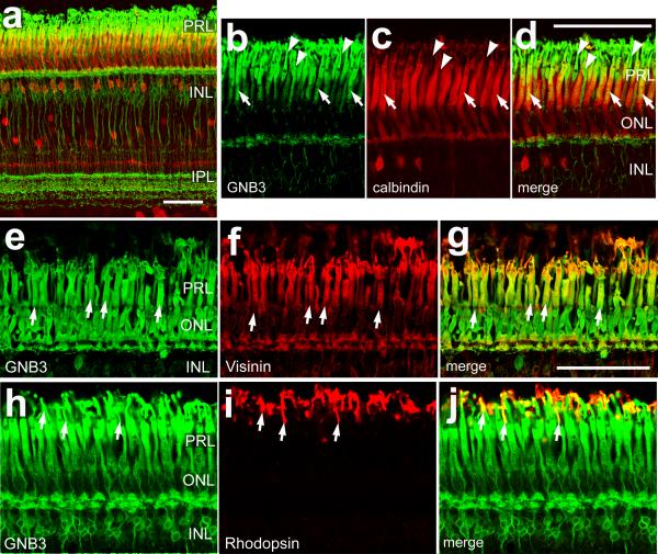
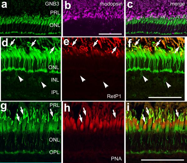
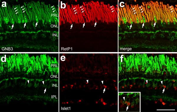
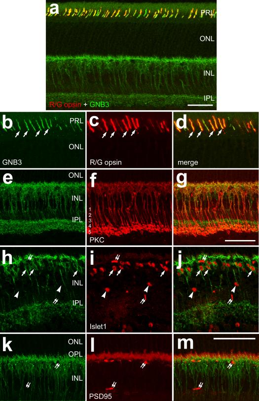
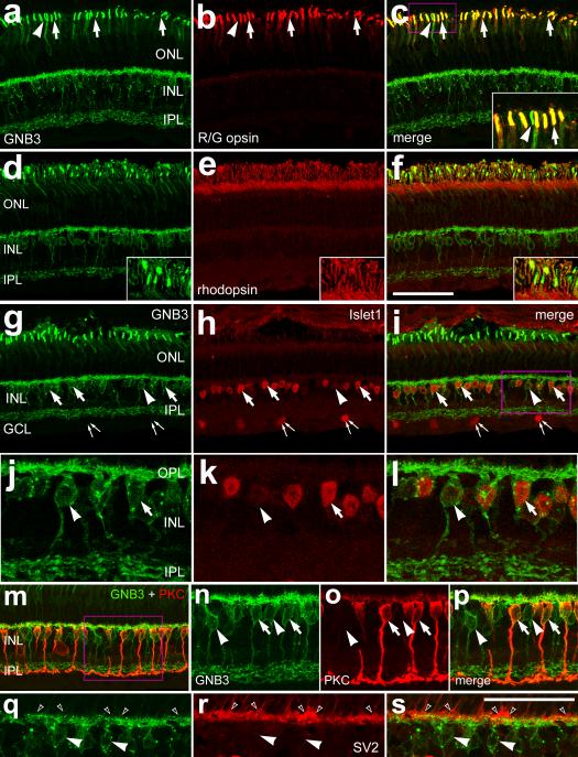
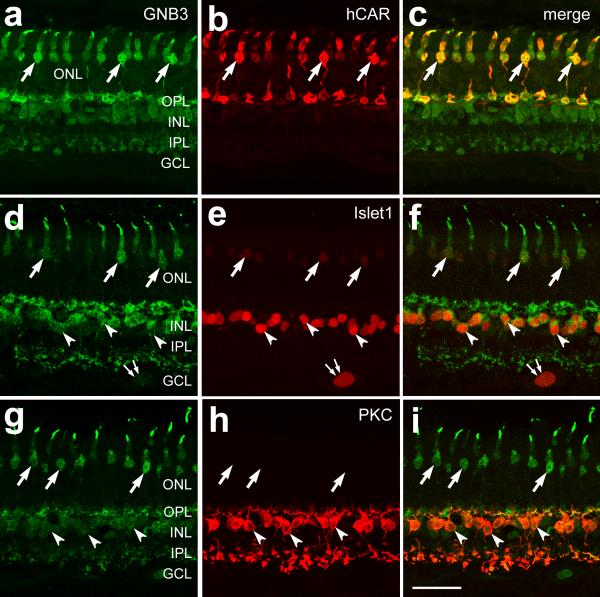
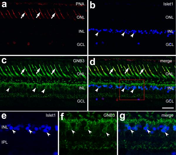
Guanine nucleotide-binding protein beta3 (GNB3) is an isoform of the beta subunit of the heterotrimeric G protein second messenger complex that is commonly associated with transmembrane receptors. The presence of GNB3 in photoreceptors, and possibly bipolar cells, has been confirmed in murine, bovine and primate retinas [Lee RH, Lieberman BS, Yamane HK, Bok D, Fung BK (1992) J Biol Chem 267:24776-24781; Peng YW, Robishaw JD, Levine MA, Yau KW (1992) Proc Natl Acad Sci U S A 89:10882-10886; Huang L, Max M, Margolskee RF, Su H, Masland RH, Euler T (2003) J Comp Neurol 455:1-10]. Studies have indicated that a mutation in the GNB3 gene causes progressive retinopathy and globe enlargement (RGE) in chickens. The goals of this study were to (1) examine the expression pattern of GNB3 in wild-type and RGE mutant chickens, (2) characterize the types of bipolar cells that express GNB3 and (3) examine whether the expression of GNB3 in the retina is conserved across vertebrate species. We find that chickens homozygous for the RGE allele completely lack GNB3 protein. We find that the pattern of expression of GNB3 in the retina is highly conserved across vertebrate species, including teleost fish (Carassius auratus), frogs (Xenopus laevis), chickens (Gallus domesticus), mice (Mus musculata), guinea-pigs (Cavia porcellus), dogs (Canis familiaris) and non-human primates (Macaca fasicularis). Regardless of the species, we find that GNB3 is expressed by Islet1-positive cone ON-bipolar cells and by cone photoreceptors. In some vertebrates, GNB3-immunoreactivity was observed in both rod and cone photoreceptors. A protein-protein alignment of GNB3 across different vertebrates, from fish to humans, indicates a high degree (>92%) of sequence conservation. Given that analogous types of retinal neurons express GNB3 in different species, we propose that the functions and the mechanisms that regulate the expression of GNB3 are highly conserved.


Figures

Figure 1
Figure 1. Expression of GNB3 mRNA and protein in wild-type and RGE -/- chickens
mRNA for GNB3 was detected in P7 wild-type and RGE -/- retinas by using RT-PCR. Each lane represents pools of retinal cDNA from different animals (a). Protein for GNB3 was detected in P7 wild-type and RGE -/- retinas by using Western blot analysis (b) and immunofluorescence(c). Vertical sections of wild-type and RGE-/- retinas were labeled with antibodies to GNB3 (c and d), Islet1, PKC or calbindin (e). Expression of bipolar cell markers (Islet1 and PKC) and the cone marker calbindin are identical at P7 in wild-type and RGE-/- retinas (e). The calibration bar (50 μm) in panel d applies to panels c, d and e. Abbreviations: RGE - retinopathy, globe enlargement, GAPDH - Glyceraldehyde 3-phosphate dehydrogenase, ONL - outer nuclear layer, INL - inner nuclear layer, IPL - inner plexiform layer, GCL - ganglion cell layer.
Figure 2
Figure 2. GNB3 expression in the developing chicken retina
Vertical sections of retina were labeled with antibodies to GNB3 (green; a-l) and Islet1 (red; b, e, h and k) and Visinin (red; c, f, i, and l). Retinas were obtained from embryos at E13 (a-c), E15 (d-f), E17 (g-i) and E18.5 (j-l). The arrowheads indicate co-labeling for GNB3 and Islet1 in bipolar cells (k). Areas outlined in yellow magnified approximately 2.5 times in the lower right corner (k and l). The calibration bar (50 μm) in panel l applies to all panels. Abbreviations: OPL - outer plexiform layer, IPL - inner plexiform layer.
Figure 3
Figure 3. GNB3 is expressed by photoreceptors and bipolar cells in the retinas of wildtype chicks
Vertical sections of P7 chicken retinas were labeled with antibodies to GNB3 (green), PKC (red, b and c), Islet1 (red, d-f) and Lim3 (red, h, i). DRAQ5 (magenta) was applied as a nuclear counter-stain. Images were obtained using confocal microscopy. Panel e’ is an approximate 3-fold magnification of the INL. Panel f is an orthogonal reconstruction of a Z-stack of optical sections through central retina labeled for Islet1 and GNB3. Arrows indicate GNB3-positive bipolar cells that are co-labeled for PKC or Islet1 (a-f). Arrowheads indicate horizontal cells that are labeled for Islet1 alone (d, f). Carets indicate bipolar cells that are GNB3-negative and intensely immunoreactive for Lim3 (g-i). Small double-arrows indicate cholinergic amacrine cells that are labeled for Islet1 (d). The calibration bar (50 μm) in panel e applies to a-c and e, the bar in i applies to g-i and the bar in d applies to d alone. Abbreviations: PRL - photoreceptor layer, ONL - outer nuclear layer, INL - inner nuclear layer, IPL - inner plexiform layer, GCL - ganglion cell layer.
Figure 4
Figure 4. GNB3 is expressed by all photoreceptors in retinas of wildtype chicken
Vertical sections of P7 chicken retinas were labeled with antibodies for GNB3 (green), calbindin (red, a, c & d), visinin (red, f and g) and rhodopsin (red, i and j). Images were obtained using confocal microscopy. Arrows indicate photoreceptors labeled for GNB3 and calbindin, visinin or rhodopsin. Arrowheads indicate photoreceptors labeled for GNB3 alone. The calibration bar (50 μm) in panel a applies to a, the bar in d applies to b-d and the bar in g applies to e-j. Abbreviations: PRL - photoreceptor layer, ONL - outer nuclear layer, INL - inner nuclear layer, IPL - inner plexiform layer.
Figure 5
Figure 5. GNB3 is expressed by cone photoreceptors and bipolar cells in the goldfish retina
Vertical sections of goldfish retinas were labeled with antibodies to GNB3 (green), rhodopsin (magenta, b and c), RetP1 (red, e and f), and PNA-lectin (red, h and i). Images were obtained using confocal microscopy. Arrows indicate photoreceptors labeled for GNB3 and RetP1 (d-f) or PNA-lectin (g-i). Arrowheads indicate bipolar cells labeled for GNB3 alone (d-f). The calibration bar (50 μm) in panel b applies to a-c, the bar in f applies to d-f and the bar in i applies to g-i. Abbreviations: PRL - photoreceptor layer, ONL - outer nuclear layer, INL - inner nuclear layer, IPL - inner plexiform layer.
Figure 6
Figure 6. GNB3 is expressed by photoreceptors and bipolar cells in the Xenopus retina
Vertical sections of Xenopus retinas were labeled with antibodies to GNB3 (green), RetP1 (red, b and c) and Islet1 (red, e and f). Small triple arrows indicate the outer segments of rod photoreceptors (a-c). Arrows in panels a-c indicate cone photoreceptors, and arrows in panels d-f indicate bipolar cells labeled for Islet 1 and GNB3. Arrowheads indicate bipolar cells labeled for GNB3 alone (d-f). The inset in panel f is enlarged approximately 3-fold. The calibration bar (50 μm) in panel f applies to a-f. Abbreviations: PRL - photoreceptor layer, ONL - outer nuclear layer, INL - inner nuclear layer, IPL - inner plexiform layer.
Figure 7
Figure 7. GNB3 is expressed by cone photoreceptors and bipolar cells in the mouse retina
Vertical sections of mouse retinas were labeled with antibodies to GNB3 (green), red/ green opsin (red, a, c and d), PKC (red, f and g), Islet1 (red, i and j) and PSD-95 (red, l and m). Arrows indicate cone photoreceptors labeled for GNB3 and red/ green opsin (b-d) or bipolar cells labeled for GNB3 and Islet1 (h-j). Arrowheads indicate cholinergic amacrine cells labeled for Islet1 (h-j). Small double arrows indicate capillaries within the retina that are labeled with secondary alone (h-m). The calibration bar (50 μm) in panel (g) applies to (b-g), the bar in (m) applies to (h-m) and the bar in (a) applies to (a) alone. Abbreviations: PRL - photoreceptor layer, ONL - outer nuclear layer, INL - inner nuclear layer, IPL - inner plexiform layer.
Figure 8
Figure 8. GNB3 is expressed by cone photoreceptors and bipolar cells in the guinea pig retina
GNB3 immunoreactivity is found in the OPL, IPL, bipolar cells and photoreceptors. Vertical sections of guinea pig retinas were labeled with antibodies for GNB3 (green), red/ green opsin (red, b and c), rhodopsin (red, e and f), Islet1 (red, h-l), PKC (red, m-p) and SV2 (red, r and s). Arrows in panels a-c indicate cones double positive for red/ green opsin and GNB3, whereas arrowheads point to GNB3 positive, red/ green opsin negative cones. Insets in panels c-f are enlarged approximately 3-fold. The areas boxed-out (magenta) in panels i and m are enlarged approximately 2-fold in panels j-l and n-p, respectively. Small double arrows indicate Islet1-positive, GNB3-negative ganglion cells (g-i). Arrows in panels g-l indicate bipolar cells that are labeled for Islet1 and GNB3, and the arrowheads indicate GNB3 positive bipolar cells. Hollow arrowheads (q-s) point to the presynaptic terminals of photoreceptors in the OPL. The calibration bar (50 μm) in panel f applies to panels a-i, bar in s applies to panels q-s. Abbreviations: ONL - outer nuclear layer; INL - inner nuclear layer; IPL - inner plexiform layer, GCL - ganglion cell layer.
Figure 9
Figure 9. GNB3 is expressed by cone photoreceptors and bipolar cells in the dog retina
Vertical sections of canine retinas were labeled with antibodies to GNB3 (green), human cone arrestin (hCAR; red, b and c), Islet1 (red, e and f) and PKC (red, h and i). Arrows indicate cone photoreceptors labeled for GNB3 and hCAR (a-c) or Islet1 (d-f), arrowheads indicate bipolar cells labeled for GNB3 and Islet1 (d-f) or PKC (g-i). The calibration bar (50 μm) in panel i applies to panels a-i. Small double arrows (d-f) indicate Islet1 positive ganglion cells. Abbreviations: ONL - outer nuclear layer, OPL - outer plexiform layer, INL - inner nuclear layer, IPL - inner plexiform layer, GCL - ganglion cell layer.
Figure 10
Figure 10. GNB3 is expressed by cone photoreceptors and bipolar cells in the primate retina (Macaca fascicularis)
Vertical sections of retina were labeled with antibodies to PNA-Lectin (red; a and d), GNB3 (green; c, d, f and g) and Islet1 (blue; b, d and e). The large arrows indicate co-labeling for GNB3 and PNA-568 in cone outer segments. The arrow heads indicate co-labeling for GNB3 and Islet1 in bipolar cells. The calibration bar (50 μm) in d applies to panels a-d. Panels e-g are enlarged approximately 3-fold. Abbreviations: ONL - outer nuclear layer; INL - inner nuclear layer, IPL - inner plexiform layer , GCL - ganglion cell layer.

Similar articles

Cited by

References

    1. Aartsen WM, Kantardzhieva A, Klooster J, van Rossum AG, van de Pavert SA, Versteeg I, Cardozo BN, Tonagel F, Beck SC, Tanimoto N, Seeliger MW, Wijnholds J. Mpp4 recruits Psd95 and Veli3 towards the photoreceptor synapse. Hum Mol Genet. 2006;15:1291–1302. - PubMed
    1. Blanks JC, Johnson LV. Specific binding of peanut lectin to a class of retinal photoreceptor cells. A species comparison. Invest Ophthalmol Vis Sci. 1984;25:546–557. - PubMed
    1. Brann MR, Cohen LV. Diurnal expression of transducin mRNA and translocation of transducin in rods of rat retina. Science. 1987;235:585–587. - PubMed
    1. Bruhn SL, Cepko CL. Development of the pattern of photoreceptors in the chick retina. J Neurosci. 1996;16:1430–1439. - PMC - PubMed
    1. Cabrera-Vera TM, Vanhauwe J, Thomas TO, Medkova M, Preininger A, Mazzoni MR, Hamm HE. Insights into G protein structure, function, and regulation. Endocr Rev. 2003;24:765–781. - PubMed

Publication types

Substances